Contents Yves Clermont, Michael Lalli, Zsuzsanna Bencsath-Makkai
LIGHT-MICROSCOPIC HISTOLOGY ATLAS
     4. Nerve Tissue
  1     2     3     4     5     6     7     8     9     10     11     12     13     14     15     16     17     18     19     20     21     22     23     24     25  
  26     27     28     29     30     31     32     33     34     35  

    4.8   Ganglion Version française
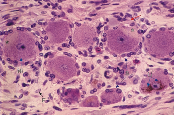
Section of a spinal ganglion.

In a few ganglion cells the axon hillocks (*), at the site of emergence of the axons, are recognized owing to the absence of Nissl bodies. The continuity of the axon hillock with the axon of a ganglion cell is indicated (red arrow).

The following structures are also labelled:

  • Basophilic Nissl bodies (Ni)
  • Satellite cells (S)
  • Lipofuscin granules (Li)
  • Fibrocyte (F) in the connective tissue

Stain: H–­­E
Magnification: ×800


We'd very much appreciate receiving your comments and suggestions!We'd very much appreciate receiving your comments and suggestions!

Creative Commons Licence The text and images of this Histology Atlas, by Yves Clermont, Michael Lalli & Zsuzsanna Bencsath-Makkai, are licensed under a Creative Commons Attribution-Noncommercial-No Derivative Works 2.5 Canada Licence and cannot be modified without the written permission of the authors. Use of any text or images must carry an acknowledgement which includes a link to the original work.


HAweb/Zs B-M -> HAbase-generated on 2013-09-13