|
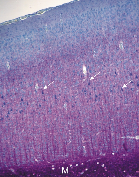
Section of a rodent cerebral cortex stained red with acid fuchsin to show the myelin of the medulla below and the myelin of the cortex above.
Note that the cortex also stains blue, but there is a progressive increase of red myelinated nerve fibres as the neurons approach the medulla (M). The latter is intensely stained red owing to the abundant myelin.
The zonation of the cortex is obvious with the large blue-stained cell bodies of pyramidal cells occupying an intermediate layer (arrows).
Stain: Toluidine Blue - Acid Fuchsin
Magnification: ×120
|