Contents Yves Clermont, Michael Lalli, Zsuzsanna Bencsath-Makkai
LIGHT-MICROSCOPIC HISTOLOGY ATLAS
     4. Nerve Tissue
  1     2     3     4     5     6     7     8     9     10     11     12     13     14     15     16     17     18     19     20     21     22     23     24     25  
  26     27     28     29     30     31     32     33     34     35  

    4.29   Cerebellum Version française
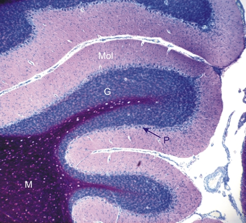
Section of the cerebellum of a rodent stained blue to show the cortex and magenta-red to show the myelin-rich medulla (M).

The cortex shows two major layers: the lightly stained superficial molecular layer (Mol) and the intensely stained subjacent basophilic granular layer (G). At the borderline of these two layers a more discreet layer is composed of the cell bodies of large neurones, the Purkinje cells (P).

Intensely stained magenta-red, the medulla (M) contains numerous myelinated axons.

Stain: Toluidine Blue - Acid Fuchsin
Magnification: ×120


We'd very much appreciate receiving your comments and suggestions!We'd very much appreciate receiving your comments and suggestions!

Creative Commons Licence The text and images of this Histology Atlas, by Yves Clermont, Michael Lalli & Zsuzsanna Bencsath-Makkai, are licensed under a Creative Commons Attribution-Noncommercial-No Derivative Works 2.5 Canada Licence and cannot be modified without the written permission of the authors. Use of any text or images must carry an acknowledgement which includes a link to the original work.


HAweb/Zs B-M -> HAbase-generated on 2013-09-13