Contents Yves Clermont, Michael Lalli, Zsuzsanna Bencsath-Makkai
LIGHT-MICROSCOPIC HISTOLOGY ATLAS
     4. Nerve Tissue
  1     2     3     4     5     6     7     8     9     10     11     12     13     14     15     16     17     18     19     20     21     22     23     24     25  
  26     27     28     29     30     31     32     33     34     35  

    4.16   Nerve Version française
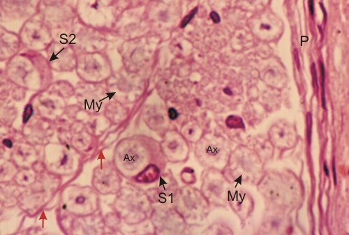
Section of a small portion of a sciatic nerve.

The connective tissue, which composes the perineurium (P) and the endoneurium (red arrows) are indicated. The slightly basophilic axons (Ax) and the partly extracted myelin sheath (My) of the nerve fibres are distinct.

Two of the fibres show the thickened cytoplasm of Schwann cells, one with a nucleus (S1) and the other without (S2). The thin cytoplasm of Schwann cells covers the nerve fibres and delimits their boundaries.

Stain: H–­­E
Magnification: ×1000


We'd very much appreciate receiving your comments and suggestions!We'd very much appreciate receiving your comments and suggestions!

Creative Commons Licence The text and images of this Histology Atlas, by Yves Clermont, Michael Lalli & Zsuzsanna Bencsath-Makkai, are licensed under a Creative Commons Attribution-Noncommercial-No Derivative Works 2.5 Canada Licence and cannot be modified without the written permission of the authors. Use of any text or images must carry an acknowledgement which includes a link to the original work.


HAweb/Zs B-M -> HAbase-generated on 2013-09-13