Contents Yves Clermont, Michael Lalli, Zsuzsanna Bencsath-Makkai
LIGHT-MICROSCOPIC HISTOLOGY ATLAS
     4. Nerve Tissue
  1     2     3     4     5     6     7     8     9     10     11     12     13     14     15     16     17     18     19     20     21     22     23     24     25  
  26     27     28     29     30     31     32     33     34     35  

    4.31   Purkinje cell Version française
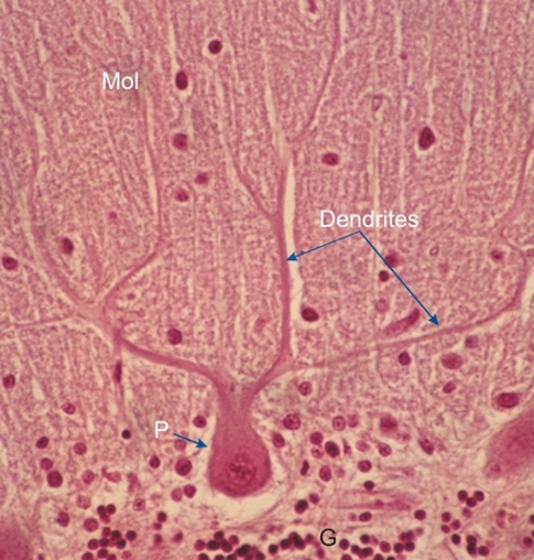
Section of the cerebellar cortex, showing the cell body of a Purkinje cell (P) at the borderline of the molecular layer (Mol) above and the granular layer (G) below.

The Purkinje cell sends numerous fanlike branching dendrites that reach the surface of the cerebellum. The single long axon of these cells (not seen here) penetrates the granular layer and the medulla.

The small nuclei present in the molecular and granular layers belong to small neurons and neuroglia.

Stain: H–­­E
Magnification: ×900


We'd very much appreciate receiving your comments and suggestions!We'd very much appreciate receiving your comments and suggestions!

Creative Commons Licence The text and images of this Histology Atlas, by Yves Clermont, Michael Lalli & Zsuzsanna Bencsath-Makkai, are licensed under a Creative Commons Attribution-Noncommercial-No Derivative Works 2.5 Canada Licence and cannot be modified without the written permission of the authors. Use of any text or images must carry an acknowledgement which includes a link to the original work.


HAweb/Zs B-M -> HAbase-generated on 2013-09-13