|
||
| 4. Nerve Tissue | ||
| 1 2 3 4 5 6 7 8 9 10 11 12 13 14 15 16 17 18 19 20 21 22 23 24 25 | ||
| 26 27 28 29 30 31 32 33 34 35 |
| |||
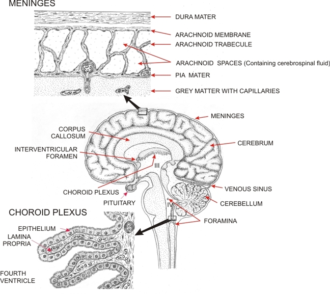 |
Drawing showing the sites of formation of the cerebrospinal fluid and its path of circulation in the central nervous system.
The cerebrospinal fluid is formed at the level of the choroid plexus. These choroid plexuses are tufts of well-vascularized villous projections attached to the wall of the four ventricles. Two of these ventricles - the 3rd (III) and the 4th (IV) - are illustrated here. The two lateral ventricles are continuous with the 3rd ventricle via the interventricular foramina. The choroid plexuses (bottom left) are lined by a simple cuboidal epithelium. These cells are instrumental in transporting fluid from the underlying porous capillaries to the lumina of the ventricles. The cerebrospinal fluid leaves the ventricles through the foramina present in the wall of the 4th ventricle (IV), enters the arachnoid spaces of the meninges covering the cebellum, and reaches these spaces at the surface of the cerebrum (top left) and of the spinal cord. The cerebrospinal fluid re-enters the venous circulation at the level of the arachnoid villi which invaginate the wall of the superior sagittal sinus. These arachnoid villi are not shown in this drawing.
|
||