Contents Yves Clermont, Michael Lalli, Zsuzsanna Bencsath-Makkai
LIGHT-MICROSCOPIC HISTOLOGY ATLAS
     4. Nerve Tissue
  1     2     3     4     5     6     7     8     9     10     11     12     13     14     15     16     17     18     19     20     21     22     23     24     25  
  26     27     28     29     30     31     32     33     34     35  

    4.2   Neuron Version française
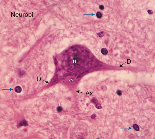
Section of the grey matter of the spinal cord. This field shows the cell body of a large neuron surrounded by the nuclei of neuroglia (arrows).

The fibrous tissue seen around the cells, called the neuropil, is composed of intertwined axons and dendrites of neurons and processes of neuroglia.

In the neuron the large nucleus (N) is surrounded by the abundant cytoplasm or perikaryon containing the basophilic Nissl bodies.

The axon hillock (*) and the axon (Ax), free of Nissl bodies, and large dendrites (D) are visible in this field

Stain: H–­­E
Magnification: ×900


We'd very much appreciate receiving your comments and suggestions!We'd very much appreciate receiving your comments and suggestions!

Creative Commons Licence The text and images of this Histology Atlas, by Yves Clermont, Michael Lalli & Zsuzsanna Bencsath-Makkai, are licensed under a Creative Commons Attribution-Noncommercial-No Derivative Works 2.5 Canada Licence and cannot be modified without the written permission of the authors. Use of any text or images must carry an acknowledgement which includes a link to the original work.


HAweb/Zs B-M -> HAbase-generated on 2013-09-13