|
||
| 4. Nerve Tissue | ||
| 1 2 3 4 5 6 7 8 9 10 11 12 13 14 15 16 17 18 19 20 21 22 23 24 25 | ||
| 26 27 28 29 30 31 32 33 34 35 |
| |||
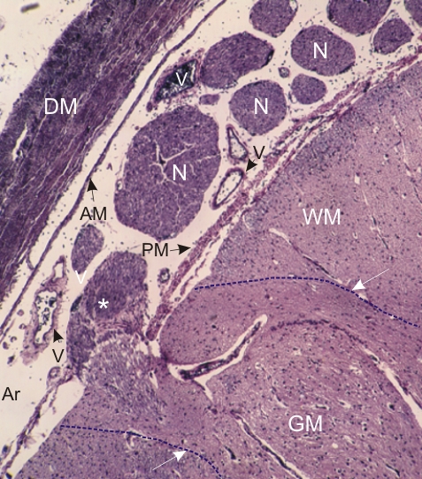 |
Section at the edge of a spinal cord and of the surrounding meninges. The tissue of the spinal cord shows the white matter (WM) and the grey matter (GM). The borderlines of these areas are indicated by stippled lines. The grey matter is continuous with a nerve rootlet (*). Several other nerves (N) are seen in the arachnoid space (Ar) of the meninges (see Figure 4.34) located between the pia mater (PM) and the arachnoid membrane (AM). The arachnoid trabecules are not distinct. Some blood vessels (V) are also present in the meninges. The dura mater (DM) surrounding the spinal cord is also labelled. Stain: HE
|
||