|
||
| 4. Nerve Tissue | ||
| 1 2 3 4 5 6 7 8 9 10 11 12 13 14 15 16 17 18 19 20 21 22 23 24 25 | ||
| 26 27 28 29 30 31 32 33 34 35 |
| |||
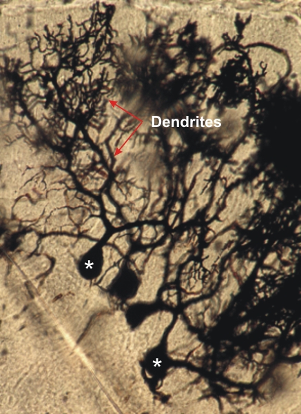 |
Thick section of a rodent cerebellum stained with silver to show the cell bodies (*) and dendrites of Purkinje cells.
The large dendrites branch out into many small dendrites that reach the surface of the molecular layer of the cortex. The axons of the Purkinje cells are not visible in this preparation. Stain: Silver
|
||