Contents Yves Clermont, Michael Lalli, Zsuzsanna Bencsath-Makkai
LIGHT-MICROSCOPIC HISTOLOGY ATLAS
     4. Nerve Tissue
  1     2     3     4     5     6     7     8     9     10     11     12     13     14     15     16     17     18     19     20     21     22     23     24     25  
  26     27     28     29     30     31     32     33     34     35  

    4.21   Nerve terminal Version française
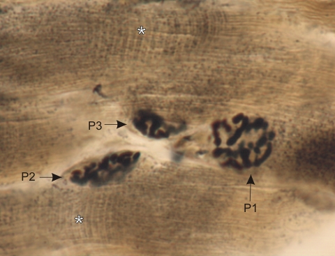
Longitudinal section of a skeletal muscle stained with silver to show motor end-plates at the surface of striated muscle fibres (*).

These nerve terminals are formed of several tortuous short branches closely applied at the surface of the muscle fibres within shallow trenches or clefts. One of these end-plates is seen in face view (P1). Two others (P2 and P3) are cut obliquely.

The lightly stained cross striations (*) of the myofibrils of muscle fibres can be identified.

Stain: Silver
Magnification: ×600


We'd very much appreciate receiving your comments and suggestions!We'd very much appreciate receiving your comments and suggestions!

Creative Commons Licence The text and images of this Histology Atlas, by Yves Clermont, Michael Lalli & Zsuzsanna Bencsath-Makkai, are licensed under a Creative Commons Attribution-Noncommercial-No Derivative Works 2.5 Canada Licence and cannot be modified without the written permission of the authors. Use of any text or images must carry an acknowledgement which includes a link to the original work.


HAweb/Zs B-M -> HAbase-generated on 2013-09-13