Contents Yves Clermont, Michael Lalli, Zsuzsanna Bencsath-Makkai
LIGHT-MICROSCOPIC HISTOLOGY ATLAS
     4. Nerve Tissue
  1     2     3     4     5     6     7     8     9     10     11     12     13     14     15     16     17     18     19     20     21     22     23     24     25  
  26     27     28     29     30     31     32     33     34     35  

    4.35   Choroid plexus Version française
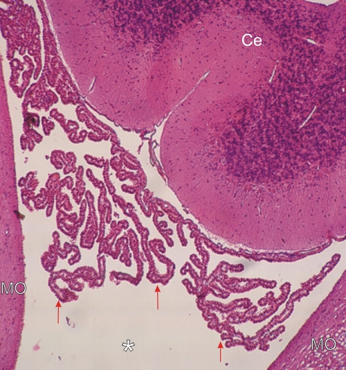
Longitudinal section through the 4th ventricle (*) delimited by the medulla oblongata (MO) and the cerebellum (Ce).

In the lumen of the ventricle the numerous projections of the choroid plexus (arrows) are present and show their simple cuboidal epithelium lining a thin core of loose connective tissue.

Well provided with small vessels, the choroid plexus is a site of production of the cerebrospinal fluid.

Stain: H–­­E
Magnification: ×125


We'd very much appreciate receiving your comments and suggestions!We'd very much appreciate receiving your comments and suggestions!

Creative Commons Licence The text and images of this Histology Atlas, by Yves Clermont, Michael Lalli & Zsuzsanna Bencsath-Makkai, are licensed under a Creative Commons Attribution-Noncommercial-No Derivative Works 2.5 Canada Licence and cannot be modified without the written permission of the authors. Use of any text or images must carry an acknowledgement which includes a link to the original work.


HAweb/Zs B-M -> HAbase-generated on 2013-09-13