|
||
| 4. Nerve Tissue | ||
| 1 2 3 4 5 6 7 8 9 10 11 12 13 14 15 16 17 18 19 20 21 22 23 24 25 | ||
| 26 27 28 29 30 31 32 33 34 35 |
| |||
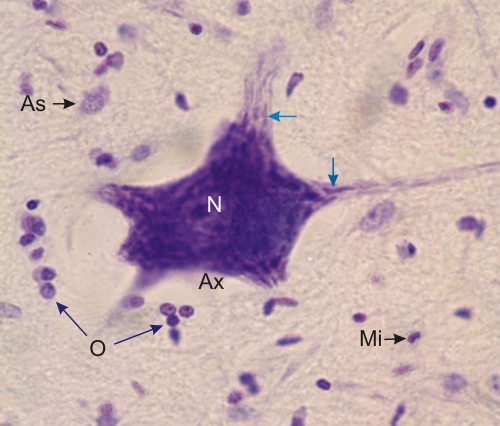 |
Section of the grey matter of a spinal cord stained with toluidine blue.
This field shows the cell body of a large motor neuron surrounded by the nuclei of several neuroglia cells. Among the latter the following cells can be identified: an astrocyte (As), oligodendrocytes (O) and microglia (Mi). In the cell body of the neurone the large central nucleus (N) is overshadowed by the basophilic Nissl bodies. These Nissl bodies are more easily seen in the dendrites (arrows). The lightly stained axon hillock at the site of emergence of the axon (Ax) is free of Nissl bodies Stain: Toluidine blue
|
||