|
||
| 4. Nerve Tissue | ||
| 1 2 3 4 5 6 7 8 9 10 11 12 13 14 15 16 17 18 19 20 21 22 23 24 25 | ||
| 26 27 28 29 30 31 32 33 34 35 |
| |||
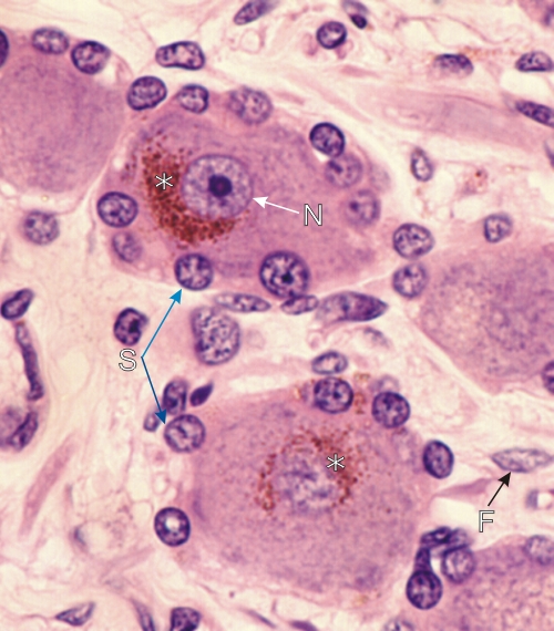 |
Section of a spinal ganglion.
This field shows two neurones: one above with a large spherical nucleus and a central nucleolus, the other one below showing a fragment of a tangentially cut nucleus without its nucleolus. Both cells contain clusters of bronze-red lipofuscin pigment granules (*). The satellite cells (S) present a spherical nucleus in an acidophilic cytoplasm. These satellite cells completely cover the body of the ganglion cells. A fibrocyte (F) is indicated in the surrounding connective tissue. Stain: HE
|
||