Contents Yves Clermont, Michael Lalli, Zsuzsanna Bencsath-Makkai
LIGHT-MICROSCOPIC HISTOLOGY ATLAS
     4. Nerve Tissue
  1     2     3     4     5     6     7     8     9     10     11     12     13     14     15     16     17     18     19     20     21     22     23     24     25  
  26     27     28     29     30     31     32     33     34     35  

    4.28   Cerebral cortex Version française
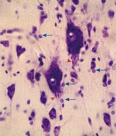
Section of a mid-layer of the cerebral cortex.

This field shows large and small pyramidal neurons and neuroglia. The intense staining of the cytoplasm of the pyramidal cells is mainly due to the basophilic Nissl bodies which are more easily seen in the dendrites (arrowheads). The nuclei of the large pyramidal cells are indicated (*).

The axons of these neurones are not stained with toluidine blue.

The small nuclei of neuroglia (arrows), most of which are oligodendrocytes are surrounded by a narrow rim of lightly stained cytoplasm.

Stain: Toluidine blue
Magnification: ×1000


We'd very much appreciate receiving your comments and suggestions!We'd very much appreciate receiving your comments and suggestions!

Creative Commons Licence The text and images of this Histology Atlas, by Yves Clermont, Michael Lalli & Zsuzsanna Bencsath-Makkai, are licensed under a Creative Commons Attribution-Noncommercial-No Derivative Works 2.5 Canada Licence and cannot be modified without the written permission of the authors. Use of any text or images must carry an acknowledgement which includes a link to the original work.


HAweb/Zs B-M -> HAbase-generated on 2013-09-13