Contents Yves Clermont, Michael Lalli, Zsuzsanna Bencsath-Makkai
LIGHT-MICROSCOPIC HISTOLOGY ATLAS
     4. Nerve Tissue
  1     2     3     4     5     6     7     8     9     10     11     12     13     14     15     16     17     18     19     20     21     22     23     24     25  
  26     27     28     29     30     31     32     33     34     35  

    4.25   Spinal cord Version française
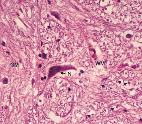
Section of a spinal cord. In this preparation the myelin of the nerve fibres was extracted by the histological procedure creating numerous artefactual spaces.

Nevertheless, the grey matter (GM) on the left and the white matter (WM) on the right can be distinguished and their approximate boundary is indicated by a broken white line. The white matter is characterized by cross sections of myelinated nerve fibres with visible axons (black arrows). The grey matter shows numerous intertwined neuronal and neuroglial processes that compose the neuropil.

The large cytoplasmic body seen at the border of the grey and white matters belongs to a large neurone (N).

Stain: H–­­E
Magnification: ×750


We'd very much appreciate receiving your comments and suggestions!We'd very much appreciate receiving your comments and suggestions!

Creative Commons Licence The text and images of this Histology Atlas, by Yves Clermont, Michael Lalli & Zsuzsanna Bencsath-Makkai, are licensed under a Creative Commons Attribution-Noncommercial-No Derivative Works 2.5 Canada Licence and cannot be modified without the written permission of the authors. Use of any text or images must carry an acknowledgement which includes a link to the original work.


HAweb/Zs B-M -> HAbase-generated on 2013-09-13