Contents Yves Clermont, Michael Lalli, Zsuzsanna Bencsath-Makkai
LIGHT-MICROSCOPIC HISTOLOGY ATLAS
     4. Nerve Tissue
  1     2     3     4     5     6     7     8     9     10     11     12     13     14     15     16     17     18     19     20     21     22     23     24     25  
  26     27     28     29     30     31     32     33     34     35  

    4.17   Nerve Version française
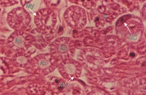
Transverse sections of sciatic nerve fibres.

Several fibres show lightly stained axons (Ax) surrounded by the partly extracted and precipitated eosinophilic myelin (My). This precipitated myelin presents a wheel-spoke configuration.

One nerve fibre shows a Schwann cell nucleus (Sc) in the crescentic cytoplasm surrounding the myelin.

A cluster of small nerve fibres (*) is indicated.

Stain: H–­­E
Magnification: ×1200


We'd very much appreciate receiving your comments and suggestions!We'd very much appreciate receiving your comments and suggestions!

Creative Commons Licence The text and images of this Histology Atlas, by Yves Clermont, Michael Lalli & Zsuzsanna Bencsath-Makkai, are licensed under a Creative Commons Attribution-Noncommercial-No Derivative Works 2.5 Canada Licence and cannot be modified without the written permission of the authors. Use of any text or images must carry an acknowledgement which includes a link to the original work.


HAweb/Zs B-M -> HAbase-generated on 2013-09-13