Contents Yves Clermont, Michael Lalli, Zsuzsanna Bencsath-Makkai
LIGHT-MICROSCOPIC HISTOLOGY ATLAS
     4. Nerve Tissue
  1     2     3     4     5     6     7     8     9     10     11     12     13     14     15     16     17     18     19     20     21     22     23     24     25  
  26     27     28     29     30     31     32     33     34     35  

    4.20   Nerve fibre Version française
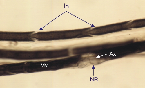
Three myelinated nerve fibres. These fibres were dissected, fixed and stained with osmic acid and mounted in toto between a glass slide and a thin cover slip.

One of these fibres shows a node of Ranvier (NR) and the traversing axon (Ax). The osmiophilic myelin, stained black, is absent at the level of this node. In addition, one fibre shows clearly oblique interruptions along the myelin sheath (My). These funnel-shaped interruptions are called incisures (In). These cone-shaped incisures, containing Schwann cell cytoplasm, have their bases located next to the axon and their mouths reaching the surface of the myelin sheath.

These incisures bridge two thin layers of Schwann cell cytoplasm: one at the surface of the myelin and the other along the axon (see Figure 4.12).

Stain: Osmium
Magnification: ×600


We'd very much appreciate receiving your comments and suggestions!We'd very much appreciate receiving your comments and suggestions!

Creative Commons Licence The text and images of this Histology Atlas, by Yves Clermont, Michael Lalli & Zsuzsanna Bencsath-Makkai, are licensed under a Creative Commons Attribution-Noncommercial-No Derivative Works 2.5 Canada Licence and cannot be modified without the written permission of the authors. Use of any text or images must carry an acknowledgement which includes a link to the original work.


HAweb/Zs B-M -> HAbase-generated on 2013-09-13