Contents Yves Clermont, Michael Lalli, Zsuzsanna Bencsath-Makkai
LIGHT-MICROSCOPIC HISTOLOGY ATLAS
     4. Nerve Tissue
  1     2     3     4     5     6     7     8     9     10     11     12     13     14     15     16     17     18     19     20     21     22     23     24     25  
  26     27     28     29     30     31     32     33     34     35  

    4.24   Spinal cord Version française
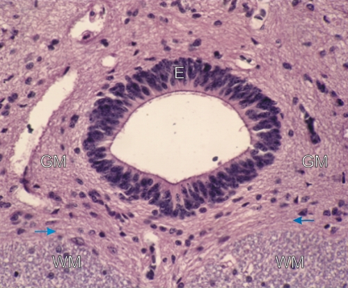
Transverse section of the central canal of the spinal cord.

The central canal is lined with a simple columnar epithelium (E) composed of neuroglial cells. These epithelial cells have a long cytoplasmic process at their base which penetrates the underlying grey matter. Such processes are not visible on this section.

The lumen of the central canal contains cerebrospinal fluid. In the surrounding nervous tissue the grey matter (GM) and the white matter (WM) can be differentiated and their borderline is indicated by arrows.

Stain: H–­­E
Magnification: ×800


We'd very much appreciate receiving your comments and suggestions!We'd very much appreciate receiving your comments and suggestions!

Creative Commons Licence The text and images of this Histology Atlas, by Yves Clermont, Michael Lalli & Zsuzsanna Bencsath-Makkai, are licensed under a Creative Commons Attribution-Noncommercial-No Derivative Works 2.5 Canada Licence and cannot be modified without the written permission of the authors. Use of any text or images must carry an acknowledgement which includes a link to the original work.


HAweb/Zs B-M -> HAbase-generated on 2013-09-13