Sommaire Yves Clermont, Michael Lalli, Zsuzsanna Bencsath-Makkai
ATLAS D'HISTOLOGIE EN MICROSCOPIE OPTIQUE
     4. Tissu Nerveux
  1     2     3     4     5     6     7     8     9     10     11     12     13     14     15     16     17     18     19     20     21     22     23     24     25  
  26     27     28     29     30     31     32     33     34     35  

    4.24   Moelle épinière English version
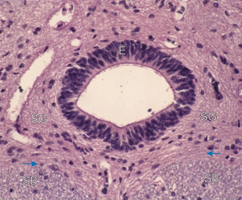
Coupe transversale du canal central de la moelle épinière.

Ce canal, qui contient le liquide céphalo-rachidien, est bordé par un épithélium simple cylindrique (E). Ses cellules épithéliales sont des cellules gliales qui possèdent un long prolongement cytoplasmique (non visible ici) qui pénètre profondément dans la substance grise sous-jacente.

Dans ce champ la substance grise (SG) est distincte de la substance blanche (SB) riche en myéline. Leur frontière est indiquée par des flèches.

Coloration: H–­­É
Grossissement: ×800


We'd very much appreciate receiving your comments and suggestions!We'd very much appreciate receiving your comments and suggestions!

Creative Commons Licence The text and images of this Histology Atlas, by Yves Clermont, Michael Lalli & Zsuzsanna Bencsath-Makkai, are licensed under a Creative Commons Attribution-Noncommercial-No Derivative Works 2.5 Canada Licence and cannot be modified without the written permission of the authors. Use of any text or images must carry an acknowledgement which includes a link to the original work.


HAweb/Zs B-M -> HAbase-generated on 2013-09-13