|
||
| 4. Tissu Nerveux | ||
| 1 2 3 4 5 6 7 8 9 10 11 12 13 14 15 16 17 18 19 20 21 22 23 24 25 | ||
| 26 27 28 29 30 31 32 33 34 35 |
| |||
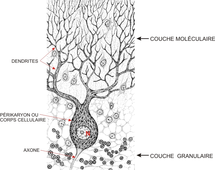 |
Ce schéma illustre une cellule de Purkinje avec son grand corps cellulaire et son grand noyau sphérique ainsi que ses innombrables dendrites qui sétendent en éventail dans la zone moléculaire.
Lunique axone de cette cellule plonge dans la zone granulaire. Les petites cellules qui entourent la cellule de Purkinje sont des petits neurones et des cellules gliales.
|
||