Sommaire Yves Clermont, Michael Lalli, Zsuzsanna Bencsath-Makkai
ATLAS D'HISTOLOGIE EN MICROSCOPIE OPTIQUE
     4. Tissu Nerveux
  1     2     3     4     5     6     7     8     9     10     11     12     13     14     15     16     17     18     19     20     21     22     23     24     25  
  26     27     28     29     30     31     32     33     34     35  

    4.5   Cellules gliales English version
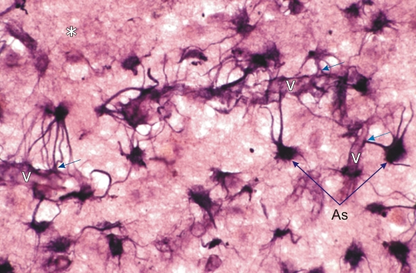
Coupe épaisse du cerveau colorée pour mettre en évidence les astrocytes (As).

Ces cellules gliales montrent de longs prolongements cytoplasmiques dont certains se terminent par des plaques (flèches) qui adhèrent à la surface des petits vaisseaux (V).

Le neuropile est marqué par un astérisque (*).

Coloration: Argent
Grossissement: ×750


We'd very much appreciate receiving your comments and suggestions!We'd very much appreciate receiving your comments and suggestions!

Creative Commons Licence The text and images of this Histology Atlas, by Yves Clermont, Michael Lalli & Zsuzsanna Bencsath-Makkai, are licensed under a Creative Commons Attribution-Noncommercial-No Derivative Works 2.5 Canada Licence and cannot be modified without the written permission of the authors. Use of any text or images must carry an acknowledgement which includes a link to the original work.


HAweb/Zs B-M -> HAbase-generated on 2013-09-13