|
||
| 4. Tissu Nerveux | ||
| 1 2 3 4 5 6 7 8 9 10 11 12 13 14 15 16 17 18 19 20 21 22 23 24 25 | ||
| 26 27 28 29 30 31 32 33 34 35 |
| |||
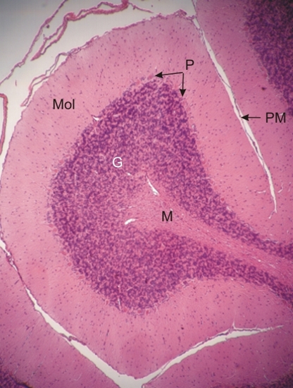 |
Coupe dun pli du cervelet. Les profondes invaginations du cortex du cervelet montre à sa surface la pie-mère (PM) des méninges. Le cortex montre la zone moléculaire (Mol), la zone granulaire (G) et à la frontière de ces deux zones, la couche de cellules de Purkinje (P). La médullaire (M) composée de substance blanche, ici peu colorée, occupe le centre du pli du cervelet. Coloration: HÉ
|
||