Sommaire Yves Clermont, Michael Lalli, Zsuzsanna Bencsath-Makkai
ATLAS D'HISTOLOGIE EN MICROSCOPIE OPTIQUE
     4. Tissu Nerveux
  1     2     3     4     5     6     7     8     9     10     11     12     13     14     15     16     17     18     19     20     21     22     23     24     25  
  26     27     28     29     30     31     32     33     34     35  
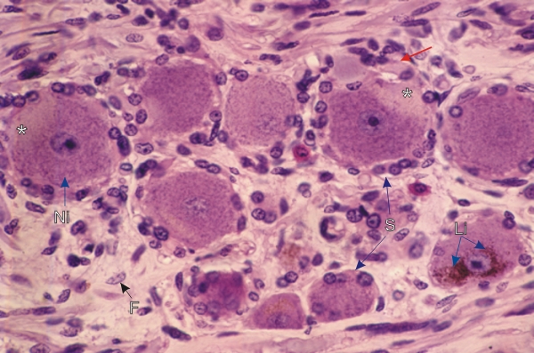
    4.8   Ganglion English version
Coupe d’un ganglion spinal.

Quelques cellules ganglionnaires montrent un cône d’implantation (*), caractérisé par l’absence de corps de Nissl. L’axone d’un de ces neurones (flèche rouge) est en continuité avec son cône d’implantation.

Les structures suivantes sont également identifiées:

  • Corps de Nissl (Ni)
  • Cellules satellites (S)
  • Granules de lipofuscine (Li)
  • Fibrocyte (F) dans le tissu conjonctif

Coloration: H–­­É
Grossissement: ×800


We'd very much appreciate receiving your comments and suggestions!We'd very much appreciate receiving your comments and suggestions!

Creative Commons Licence The text and images of this Histology Atlas, by Yves Clermont, Michael Lalli & Zsuzsanna Bencsath-Makkai, are licensed under a Creative Commons Attribution-Noncommercial-No Derivative Works 2.5 Canada Licence and cannot be modified without the written permission of the authors. Use of any text or images must carry an acknowledgement which includes a link to the original work.


HAweb/Zs B-M -> HAbase-generated on 2013-09-13