|
||
| 4. Nerve Tissue | ||
| 1 2 3 4 5 6 7 8 9 10 11 12 13 14 15 16 17 18 19 20 21 22 23 24 25 | ||
| 26 27 28 29 30 31 32 33 34 35 |
| |||
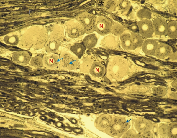 |
Longitudinal section of a mouse cervical ganglion stained with osmium to show the cell bodies of ganglion cells surrounded by osmiophilic myelinated nerve fibres (F).
In the cytoplasm surrounding the unstained nuclei (N) of some ganglion cells, the osmiophilic bodies (arrows) belong to the Golgi apparatus (see details in Figure 4.10). The osmiophilic myelinated fibres (F) are seen in longitudinal, oblique and cross sections. (N.B.: The osmiophilic bodies present in the perikaryon of such ganglion cells were first discovered by Golgi in 1898!) Stain: Osmium
|
||