 |
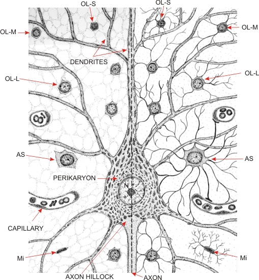
The panel on the left shows the neuron and the neuroglia as seen in a standard histological section. The panel on the right illustrates the neuroglia with their cytoplasmic processes.
The cell body of the neuron has a large spherical nucleus (N) surrounded by an abundant cytoplasm called the perikaryon. It contains the Nissl bodies composed of ER cisternae associated with ribosomes. The spaces between the Nissl bodies are occupied by ribosome-free ER cisternae, the Golgi apparatus, mitochondria, neurofilaments and neurotubules (not illustrated in this drawing). At the base of the neuron, at the region of the axon hillock, the perikaryon is continuous with the axon. Both axon hillock and axon are free of Nissl bodies and of the Golgi apparatus. Numerous branching dendrites extend around and above the perikaryon. These dendrites contain Nissl bodies, mitochondria, filaments and tubules but no Golgi apparatus.
Surrounding the neuron are several neuroglia and some capillaries. On the left, these glial cells have small nuclei surrounded by a thin rim of cytoplasm. On the right, their stellate cytoplasmic processes are represented. Such processes surround and insulate the cell body, axon and dendrites (except the synapses which are points of contact between neurons). There are three categories of neuroglia in the grey matter: the oligodendrocytes (OL) are the most numerous and show spherical nuclei of various sizes; the astrocytes (AS) are the largest and show processes with platelike extremities applied to the surface of small blood vessels; the microglia (Mi) have a small nucleus surrounded by hairy cytoplasmic processes. The microglia of bone marrow origin can show phagocytic activity. Oligodendrocytes are labelled as follows: large (OL-L), medium (OL-M) and small (OL-S).
|