|
||
| 4. Nerve Tissue | ||
| 1 2 3 4 5 6 7 8 9 10 11 12 13 14 15 16 17 18 19 20 21 22 23 24 25 | ||
| 26 27 28 29 30 31 32 33 34 35 |
| |||
 |
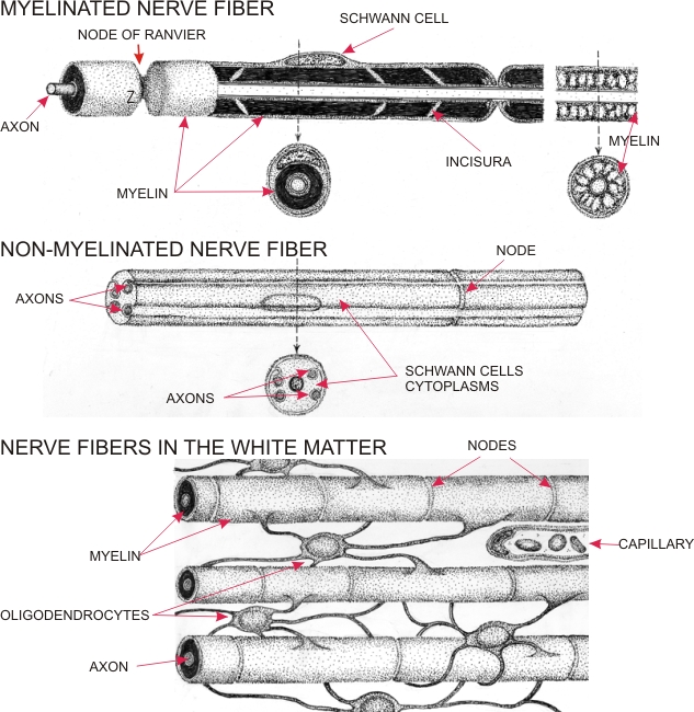
Drawing showing nerve fibres and associated neuroglia.
In the panel above, the nerve fibres of the peripheral nervous system are shown in longitudinal and cross sections. These nerves fibres are of two types: myelinated (above) and non-myelinated (below). In the myelinated fibres each axon is covered by a myelin sheath associated with Schwann cells, a class of neuroglia. The nucleus of a Schwann cell is at the surface of the fibre. Schwann cells are placed end to end. At their points of contact the myelin sheath is interrupted by constrictions called nodes of Ranvier. The myelin presents oblique interruptions, called incisures, which contain Schwann cell cytoplasm. In nerve fibres fixed and stained with a usual method, the myelin is partially extracted and the residues form irregular fibrous networks (top diagram, right). The non-myelinated fibres have a central nucleus of a Schwann cell surrounded by a cytoplasm in which one or many axons are inserted. Nodes are present at the points of contact of Schwann cells along the nerve fibre. In the panel below, the drawing illustrates nerve fibres in the white matter. These fibres are associated with oligodendrocytes responsible for depositing of myelin at the surface of axons. Note that a single oligodendrocyte sends cytoplasmic processes to several adjacent axons.
|
||