|
||
| 11. Oral Cavity | ||
| 1 2 3 4 5 6 7 8 9 10 11 12 13 14 15 16 17 18 19 20 21 22 23 24 25 | ||
| 26 27 28 29 30 31 32 33 34 35 36 37 38 39 40 41 |
| |||
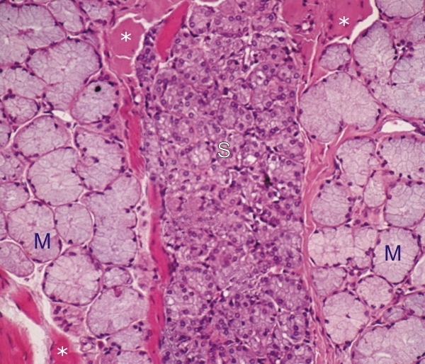 |
Section of a human tongue.
This is a side-by-side view of groups of mucous acini (M) and serous acini (S) separated by striated muscle fibres (*). The mucous acini show large glandular cells with a lightly stained cytoplasm, due to the partial extraction of water-soluble mucigen granules. Their nuclei are flattened at the base of the cytoplasm. In contrast, the smaller glandular cells of the serous acini have a more heavily stained cytoplasm and a spherical basal nucleus (see Figure 11.12.). Stain: HE
|
||