|
||
| 11. Oral Cavity | ||
| 1 2 3 4 5 6 7 8 9 10 11 12 13 14 15 16 17 18 19 20 21 22 23 24 25 | ||
| 26 27 28 29 30 31 32 33 34 35 36 37 38 39 40 41 |
| |||
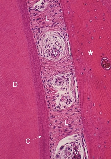 |
Root of a tooth.
This field shows the dentin (D), the bone (*) and the periodontal ligament (L) that bridges the tooth to the bone. This ligament is composed of type I collagen fibres and of numerous fibrocytes. The connective tissue fibres are inserted in both the basophilic acellular cementum (C) at the surface of the root and the bone matrix. This periodontal ligament shows an area of loose connective tissue containing small blood vessels (V). Stain: HE
|
||