Contents Yves Clermont, Michael Lalli, Zsuzsanna Bencsath-Makkai
LIGHT-MICROSCOPIC HISTOLOGY ATLAS
     11. Oral Cavity
  1     2     3     4     5     6     7     8     9     10     11     12     13     14     15     16     17     18     19     20     21     22     23     24     25  
  26     27     28     29     30     31     32     33     34     35     36     37     38     39     40     41  

    11.30   Tooth and cementum Version française
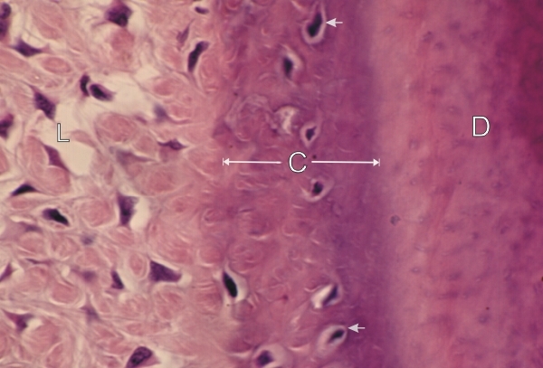
Oblique section of the root of a human tooth.

This field shows the slightly basophilic cellular cementum (C) covering the dentin (D) at the tip of the tooth close to the apical foramen of the dental canal.

This mineralized cellular cementum is similar to bone tissue and contains cementocytes (arrows) in lacunae similar to those occupied by osteocytes.

The cellular cementum serves as a site of insertion of collagen fibres of the periodontal ligament (L).

Stain: H–­­E
Magnification: ×1000


We'd very much appreciate receiving your comments and suggestions!We'd very much appreciate receiving your comments and suggestions!

Creative Commons Licence The text and images of this Histology Atlas, by Yves Clermont, Michael Lalli & Zsuzsanna Bencsath-Makkai, are licensed under a Creative Commons Attribution-Noncommercial-No Derivative Works 2.5 Canada Licence and cannot be modified without the written permission of the authors. Use of any text or images must carry an acknowledgement which includes a link to the original work.


HAweb/Zs B-M -> HAbase-generated on 2013-09-13