|
||
| 5. Vessels | ||
| 1 2 3 4 5 6 7 8 9 10 11 12 13 14 15 16 17 18 19 20 21 22 23 24 25 | ||
| 26 27 28 29 30 31 32 33 34 35 |
| |||
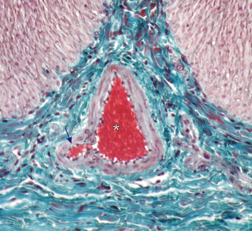 |
Section of the wall of the small intestine.
In the centre of the field, a cross section of an arteriole is filled with red blood cells (*). A smaller arteriole (arrow) branches from the larger one. The media of these vessels are composed of two or three layers of smooth muscle cells. These small vessels are surrounded by dense connective tissue, stained blue-green, in which the adventitia of the arterioles cannot be demarcated. Stain: Massons Trichrome
|
||