Contents Yves Clermont, Michael Lalli, Zsuzsanna Bencsath-Makkai
LIGHT-MICROSCOPIC HISTOLOGY ATLAS
     5. Vessels
  1     2     3     4     5     6     7     8     9     10     11     12     13     14     15     16     17     18     19     20     21     22     23     24     25  
  26     27     28     29     30     31     32     33     34     35  

    5.21   Artery and vein Version française
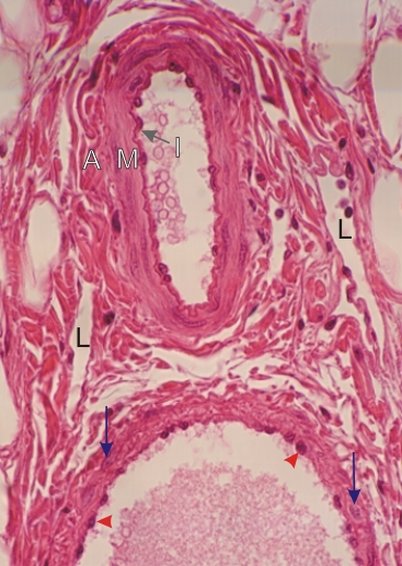
Section of a small artery (top) and of a small vein (bottom).

In the artery the endothelial cells and the internal elastic lamella form the intima (I). The media (M) shows three concentric layers of smooth muscle cells. The adventitia (A), composed of dense connective tissue, has no sharp external boundary.

In the surrounding connective tissue, some small lymphatic vessels (L) are visible.

The accompanying vein shows the endothelium (arrowheads) but no internal elastic lamella.

The wall of the vein is composed of a layer of dense connective tissue in which the nuclei of a few smooth muscle cells can be identified (blue arrows).

Stain: H–­­E
Magnification: ×600


We'd very much appreciate receiving your comments and suggestions!We'd very much appreciate receiving your comments and suggestions!

Creative Commons Licence The text and images of this Histology Atlas, by Yves Clermont, Michael Lalli & Zsuzsanna Bencsath-Makkai, are licensed under a Creative Commons Attribution-Noncommercial-No Derivative Works 2.5 Canada Licence and cannot be modified without the written permission of the authors. Use of any text or images must carry an acknowledgement which includes a link to the original work.


HAweb/Zs B-M -> HAbase-generated on 2013-09-13