|
||
| 3. Muscle Tissue | ||
| 1 2 3 4 5 6 7 8 9 10 11 12 13 14 15 16 17 |
| |||
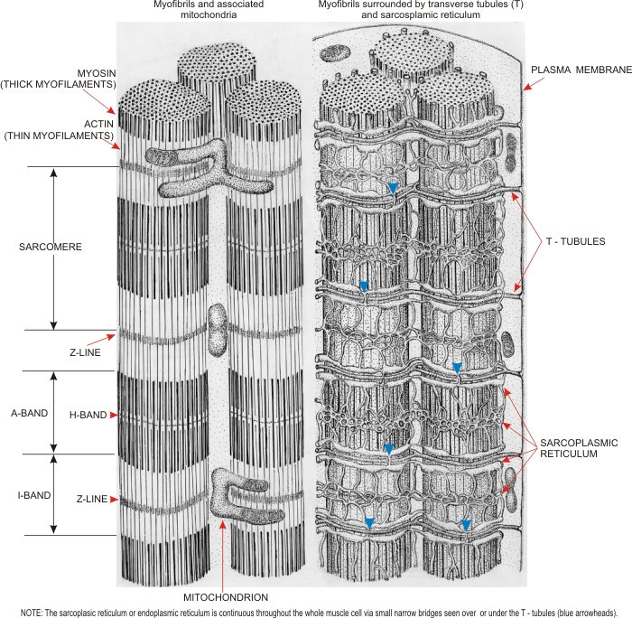 |
Drawings of myofibrils of skeletal muscle fibres of mammals derived from electron-microscope studies.
(Left) The arrangement of thin actin and thick myosin myofilaments along the A- and I-bands of the myofibrils are depicted, along with some mitochondria. ) (Right) The transverse or T-tubules are continuous with the plasma membrane and circle the myofibrils at the borderline of the A- and I-bands. This drawing also illustrates the networks of cisternae of smooth endoplasmic reticulum or sarcoplasmic reticulum which surround the myofibrils. The cisternae of the sarcoplasmic reticulum, via connections over the T-tubules (arrowheads), form a single continuous system throughout the muscle fibre. See textbooks for presentations of (a) the role of actin and myosin filaments during the contraction and relaxation of muscle fibres, and (b) the role of T-tubules and cisternae of endoplasmic reticulum during the nervous stimulation leading to the contractions of the myofibrils.
|
||