Contents Yves Clermont, Michael Lalli, Zsuzsanna Bencsath-Makkai
LIGHT-MICROSCOPIC HISTOLOGY ATLAS
     3. Muscle Tissue
  1     2     3     4     5     6     7     8     9     10     11     12     13     14     15     16     17  

    3.15   Smooth muscle fibres Version française
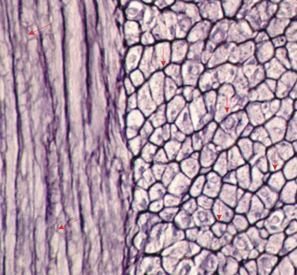
Section of the muscular coats of the small intestine stained with silver nitrate to show in black the reticular fibres (type III collagen).

On the left the smooth muscle cells are cut longitudinally and on the right they are cut transversely.

In this thick section of smooth muscle cells cut transversely, the cells appear completely surrounded by black reticular fibres (vertical arrows).

On the left the reticular fibres form networks, seen in face view (oblique arrows), at the surface of the muscle cells.

Stain: Silver
Magnification: ×1000


We'd very much appreciate receiving your comments and suggestions!We'd very much appreciate receiving your comments and suggestions!

Creative Commons Licence The text and images of this Histology Atlas, by Yves Clermont, Michael Lalli & Zsuzsanna Bencsath-Makkai, are licensed under a Creative Commons Attribution-Noncommercial-No Derivative Works 2.5 Canada Licence and cannot be modified without the written permission of the authors. Use of any text or images must carry an acknowledgement which includes a link to the original work.


HAweb/Zs B-M -> HAbase-generated on 2013-09-13