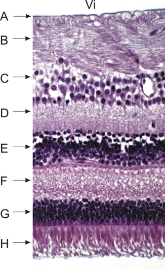
Contents Yves Clermont, Michael Lalli, Zsuzsanna Bencsath-Makkai
LIGHT-MICROSCOPIC HISTOLOGY ATLAS
     18. Eye
  1     2     3     4     5     6     7     8     9     10     11     12     13     14     15     16     17     18     19     20     21     22     23     24     25  
  26     27     28     29  

    18.28   Retina Version française
Dog’s retina at a short distance from the optic nerve.

The following structures are identified:

  1. Basement membrane at the borderline of the vitreous body (Vi) and the retina
  2. Layer of optic nerve fibres seen here in longitudinal sections
  3. Thick layer of ganglion cells
  4. Inner plexiform layer
  5. Inner nuclear layer
  6. Outer plexiform layer
  7. Outer nuclear layer
  8. Layer of rods and cones

The pigmented cell layer of the retina, artificially detached from the rods and cones, is not present in this field.

Stain: H–­­E
Magnification: ×350


We'd very much appreciate receiving your comments and suggestions!We'd very much appreciate receiving your comments and suggestions!

Creative Commons Licence The text and images of this Histology Atlas, by Yves Clermont, Michael Lalli & Zsuzsanna Bencsath-Makkai, are licensed under a Creative Commons Attribution-Noncommercial-No Derivative Works 2.5 Canada Licence and cannot be modified without the written permission of the authors. Use of any text or images must carry an acknowledgement which includes a link to the original work.


HAweb/Zs B-M -> HAbase-generated on 2013-09-13