|
||
| 18. Eye | ||
| 1 2 3 4 5 6 7 8 9 10 11 12 13 14 15 16 17 18 19 20 21 22 23 24 25 | ||
| 26 27 28 29 |
| |||
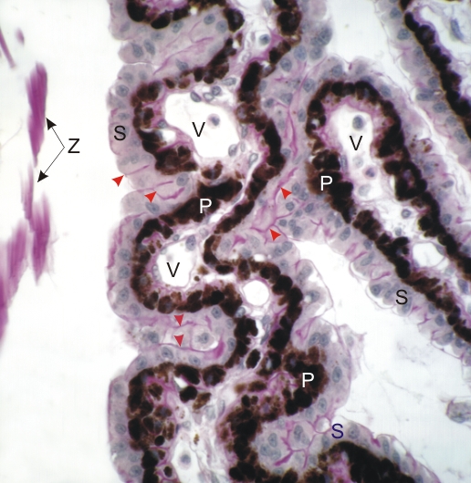 |
Sections of ciliary processes from a dogs eye stained with PAS.
The epithelium lining the branching ciliary processes shows the basal pigmented cells (P) applied to the underlying connective tissue. This tissue shows many small vessels (V). The superficial non-pigmented epithelial cells (S), facing the posterior chamber of the eye, are associated with a thin PAS-positive layer (red arrowheads) which corresponds to a basement membrane (collagen type IV). This basement membrane serves as the insertion site of zonular fibres (Z). These zonular fibres are also PAS-positive. The ciliary body and processes are sites of production of aqueous humour. Stain: PAS-Hematoxylin
|
||