Contents Yves Clermont, Michael Lalli, Zsuzsanna Bencsath-Makkai
LIGHT-MICROSCOPIC HISTOLOGY ATLAS
     1. Epithelia
  1     2     3     4     5     6     7     8     9     10     11     12     13     14     15     16     17     18     19     20     21     22     23     24  

    1.7   Simple cuboidal epithelia of urinary tubules Version française
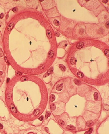
Transverse sections of two types of urinary tubules both lined with simple cuboidal epithelia.

Some tubules (*) are lined with cuboidal cells showing distinct lateral cell membranes.

The other three tubules (+) are lined with low cuboidal cells with indistinct lateral cytoplasmic membranes. In this case, the lateral cell membrane is present but forms deep lateral folds in the acidophilic cytoplasm, which renders them invisible under a light microscope.

Stain: H–­­E
Magnification: ×800


We'd very much appreciate receiving your comments and suggestions!We'd very much appreciate receiving your comments and suggestions!

Creative Commons Licence The text and images of this Histology Atlas, by Yves Clermont, Michael Lalli & Zsuzsanna Bencsath-Makkai, are licensed under a Creative Commons Attribution-Noncommercial-No Derivative Works 2.5 Canada Licence and cannot be modified without the written permission of the authors. Use of any text or images must carry an acknowledgement which includes a link to the original work.


HAweb/Zs B-M -> HAbase-generated on 2013-09-13