|
||
| 1. Epithelia | ||
| 1 2 3 4 5 6 7 8 9 10 11 12 13 14 15 16 17 18 19 20 21 22 23 24 |
| |||
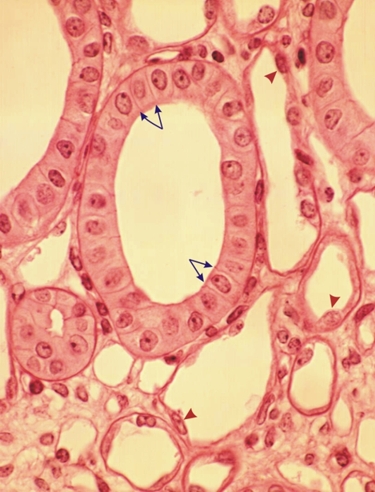 |
Section of urinary tubules from a dog kidney.
This image shows several cross and oblique sections of ducts. The large ones are lined with tall cuboidal cells showing distinct lateral cell membranes (arrows) and central ovoid nuclei. They form a typical simple cuboidal epithelium. Other ducts are lined with flat epithelial cells characteristic of simple squamous epithelia (arrowheads). Stain: HE
|
||