|
||
| 14. Glandes Endocrines | ||
| 1 2 3 4 5 6 7 8 9 10 11 12 13 14 15 16 17 18 19 20 21 22 23 24 25 | ||
| 26 27 28 29 30 |
| |||
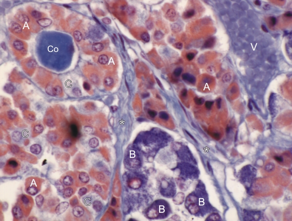 |
Coupe du lobe antérieur dune pituitaire humaine colorée au bleu daniline et à lorcéine.
Les acidophiles (A) sont colorés en rouge-orangé, les basophiles ( B) en bleu et les chromophobes (C) en bleu-pâle. Un groupe de cellules, surtout des acidophiles ici, forment un kyste avec une lumière centrale montrant une colloïde (Co) colorée en bleu. Du tissu conjonctif (*) et un petit vaisseau (V) sont également identifiés. Coloration: Bleu daniline et orcéine
|
||