|
||
| 14. Glandes Endocrines | ||
| 1 2 3 4 5 6 7 8 9 10 11 12 13 14 15 16 17 18 19 20 21 22 23 24 25 | ||
| 26 27 28 29 30 |
| |||
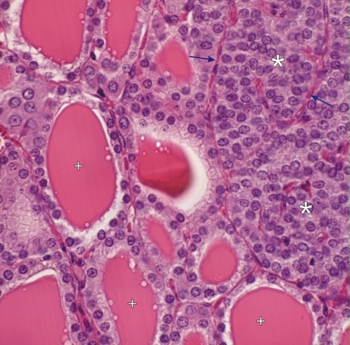 |
Glandes thyroïde et parathyroïde dun chien. La parathyroïde (à droite) est formée de groupes de petites cellules glandulaires (*) séparés par une petite quantité de tissu conjonctif (flèches). Également visibles; les cellules folliculaires des follicules et les colloïdes des follicules adjacents (+). Coloration: HÉ
|
||