Contents Yves Clermont, Michael Lalli, Zsuzsanna Bencsath-Makkai
LIGHT-MICROSCOPIC HISTOLOGY ATLAS
     13. Urinary System
  1     2     3     4     5     6     7     8     9     10     11     12     13     14     15     16     17     18     19     20     21     22     23     24     25  
  26     27  

    13.17   Kidney cortex and medulla Version française
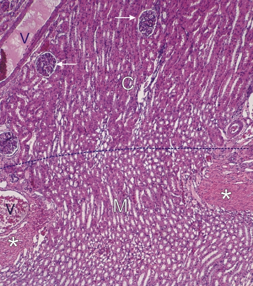
Section of the kidney showing the borderline (dotted line) of the cortex (C) and the medulla (M).

The cortex above can readily be recognized by the presence of renal corpuscles (arrows). The medulla, below the line, is free of renal corpuscles. It contains the segments of the loops of Henle seen here in transverse and longitudinal sections.

Also indicated are some dense connective tissue (*) and blood vessels (V).

Stain: H–­­E
Magnification: ×150


We'd very much appreciate receiving your comments and suggestions!We'd very much appreciate receiving your comments and suggestions!

Creative Commons Licence The text and images of this Histology Atlas, by Yves Clermont, Michael Lalli & Zsuzsanna Bencsath-Makkai, are licensed under a Creative Commons Attribution-Noncommercial-No Derivative Works 2.5 Canada Licence and cannot be modified without the written permission of the authors. Use of any text or images must carry an acknowledgement which includes a link to the original work.


HAweb/Zs B-M -> HAbase-generated on 2013-09-13