|
||
| 10. Respiratory System | ||
| 1 2 3 4 5 6 7 8 9 10 11 12 13 14 15 16 17 18 19 20 21 22 23 24 25 | ||
| 26 27 |
| |||
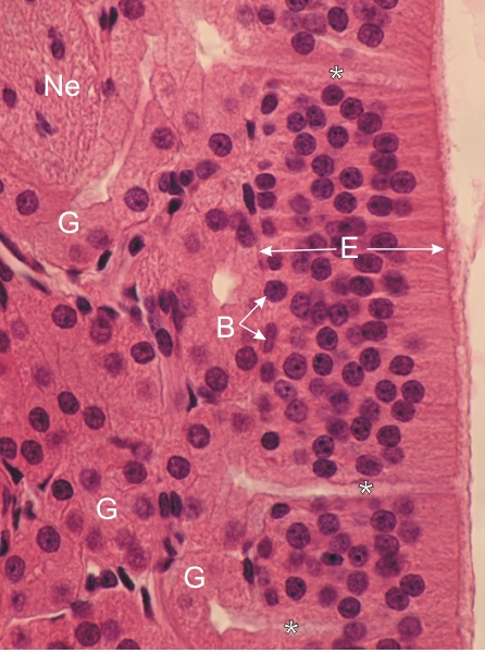 |
Section of the olfactory mucosa of a monkey. This is a higher magnification of the framed area in Figure 10.1.
The pseudostratified columnar epithelium (E) lining the mucosa is composed of basal cells (B) close to the lamina propria, and of two types of spindle-shaped cells, here indistinct from each other: the sensory olfactory neurones and the supporting cells. Both cell types have spherical nuclei located at various depths in the cytoplasm of epithelial cells. The olfactory cells have small filiform axonal processes (not visible with the light microscope) that cross the basement membrane and form non-myelinated nerves (Ne). The serous Bowmans glands (G) have small ducts (*) that cross the epithelium and open at the surface of this epithelium. Stain: HE
|
||