Contents Yves Clermont, Michael Lalli, Zsuzsanna Bencsath-Makkai
LIGHT-MICROSCOPIC HISTOLOGY ATLAS
     6. Lymphatic Organs
  1     2     3     4     5     6     7     8     9     10     11     12     13     14     15     16     17     18     19     20     21     22     23     24     25  
  26     27     28     29     30     31     32     33     34     35     36     37     38  

    6.37   Spleen Version française
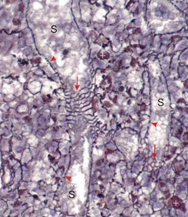
Section of a rodent spleen stained with silver to show the reticular fibres in black.

In this field the red pulp shows thick argyrophilic reticular fibres around venous sinuses. Parts of this network are seen in a face view (arrows). The rest of the network is seen in a side view (arrowheads).

Some brownish macrophages (+) are present in the loose networks of reticular fibres of the splenic cords.

Stain: Hematoxylin and silver
Magnification: ×750


We'd very much appreciate receiving your comments and suggestions!We'd very much appreciate receiving your comments and suggestions!

Creative Commons Licence The text and images of this Histology Atlas, by Yves Clermont, Michael Lalli & Zsuzsanna Bencsath-Makkai, are licensed under a Creative Commons Attribution-Noncommercial-No Derivative Works 2.5 Canada Licence and cannot be modified without the written permission of the authors. Use of any text or images must carry an acknowledgement which includes a link to the original work.


HAweb/Zs B-M -> HAbase-generated on 2013-09-13