|
||
| 6. Organes Lymphatiques | ||
| 1 2 3 4 5 6 7 8 9 10 11 12 13 14 15 16 17 18 19 20 21 22 23 24 25 | ||
| 26 27 28 29 30 31 32 33 34 35 36 37 38 |
| |||
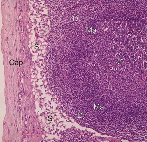 |
Coupe du cortex dun ganglion lymphatique.
Sous la capsule conjonctive (Cap), la zone pâle correspond à un sinus lymphatique sous-capsulaire (S). Le tissu sous-jacent montre un nodule lymphatique avec son centre germinatif (G) et son manteau (Ma) plus dense et chromophile. Au-delà du nodule, le tissu lymphatique correspond au cortex diffus (D). Il faut noter quil ny a pas de frontières nettes entre les diverses zones du tissu lymphatique cortical. Coloration: HÉ
|
||