Sommaire Yves Clermont, Michael Lalli, Zsuzsanna Bencsath-Makkai
ATLAS D'HISTOLOGIE EN MICROSCOPIE OPTIQUE
     18. Œil
  1     2     3     4     5     6     7     8     9     10     11     12     13     14     15     16     17     18     19     20     21     22     23     24     25  
  26     27     28     29  

    18.14   Corps ciliaire English version
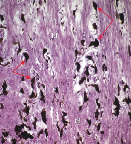
Cette coupe du corps ciliaire montre des faisceaux de fibres musculaires lisses (M) séparés par un tissu conjonctif lâche contenant de nombreux mélanocytes (flèches).

Ces faisceaux de cellules musculaires présentent deux orientations principales dans le corps ciliaire; ils sont disposés soit longitudinalement et en éventail, soit circulairement à proximité des procès ciliaires (voir la figure 18.10).

Ces muscles ciliaires jouent un rôle important dans l’accommodation visuelle.

Coloration: H–­­É
Grossissement: ×300


We'd very much appreciate receiving your comments and suggestions!We'd very much appreciate receiving your comments and suggestions!

Creative Commons Licence The text and images of this Histology Atlas, by Yves Clermont, Michael Lalli & Zsuzsanna Bencsath-Makkai, are licensed under a Creative Commons Attribution-Noncommercial-No Derivative Works 2.5 Canada Licence and cannot be modified without the written permission of the authors. Use of any text or images must carry an acknowledgement which includes a link to the original work.


HAweb/Zs B-M -> HAbase-generated on 2013-09-13