|
||
| 12. Digestive System | ||
| 1 2 3 4 5 6 7 8 9 10 11 12 13 14 15 16 17 18 19 20 21 22 23 24 25 | ||
| 26 27 28 29 30 31 32 33 34 35 36 37 38 39 40 41 42 43 44 45 46 47 48 49 50 | ||
| 51 52 53 54 55 56 57 58 59 60 61 62 63 64 65 66 67 68 69 70 71 72 73 74 75 | ||
| 76 77 78 79 80 81 82 83 84 85 86 |
| |||
 |
The liver is a large gland located in the upper region of the abdominal cavity. It weighs from 1400 to 1600 g in adult males and from 1200 to 1400 g in adult females.
The liver has two major functions:
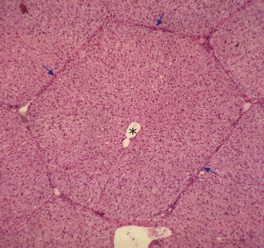
The liver, covered with a thin connective tissue membrane, is divided into a few large lobes which are subdivided into lobules. In humans and many mammalian species the demarcations of liver lobules are not clear in histological sections and the liver tissue appears homogeneous. However, in a few species, such as the pig, the liver lobules are delimited by connective tissue septae as seen in this image (arrows). Such units, polyhedral in three dimensions, are referred to as anatomical or classical lobules. Since such lobules are ill defined in humans and rodents, some histophysiologists have subdivided the liver parenchyma into functional territories (portal lobules, liver acini, acinar zones, etc.), which however cannot be easily demarcated in usual histological preparations. The present chapter will therefore be limited to descriptions of the so-called classical lobule, to the liver cells (to be called hepatocytes), to the excretory ducts and to the associated blood vessels. In this image, in the centre of the hexagonal lobule, a large vessel, the central vein (*), drains the blood from the surrounding parenchyma and carries it to the hepatic vein. In some corners of the hexagon there are vessels (i.e., portal veins, hepatic arteries) and ducts that will be examined at higher magnifications further on. Stain: HE
|
||