Middle Ear


ossicles

Tympanic Cavity


Ossicles

Malleus

malleus Malleus (VRML model)

Incus

incus Incus (VRML model)

Stapes

stapes Stapes (VRML model)

Overview of the Middle Ear

Articulation of the ossicles

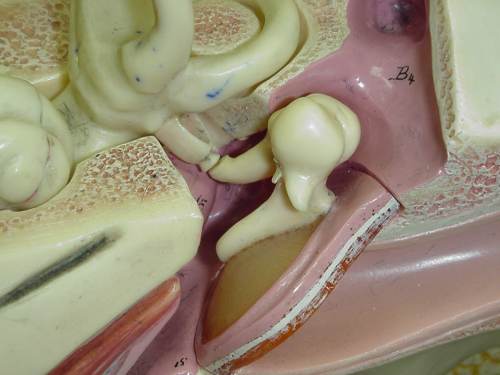
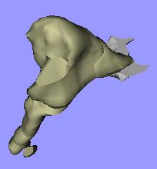
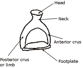
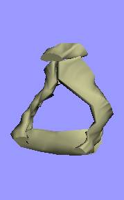
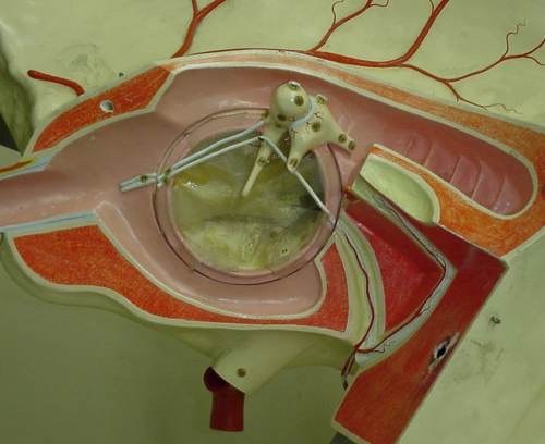
These bones form a chain across the tympanic cavity from the TM to fenestra vestibuli (oval window). Covered by mucous membrane that also lines the tympanic cavity. Transmits vibration movements of the TM to the inner ear. Functions to increase the force but decrease the amplitude. Ossicles (VRML model)
Ossicles, muscles, ligaments and TM (VRML model)
  • White: tympanic membrane and annular ligament, and posterior incudal ligament
  • Red: stapedius muscle and tensor tympani


Nerves in the middle ear

Middle and inner ear (AVI movie)
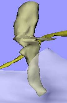
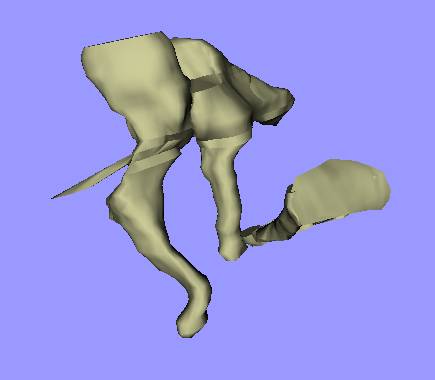
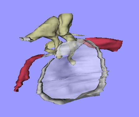
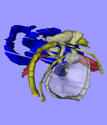
White: tympanic membrane and ligaments
Cream: malleus, incus, stapes
Red: stapedius muscle and tensor tympani
Blue: inner ear
Yellow: nerves


Schematic view of the middle-ear cavity

Lateral wall (the tympanic membrane) has been removed to see the interior of the middle ear.
  • RED: Internal carotid
  • BLUE: Internal jugular vein
  • PURPLE: Tensor tympani
  • YELLOW: Facial nerve(VII) with nerve to stapedius & chorda tympani
  • DARK GREEN: Auditory tube, oval and round windows
  • LIGHT GREEN: Cochlea
Middle ear (tympanic cavity)

Home page
Previous page
Next page
Questions?, comments,...
Last modified: Thu, 2001 May 03 15:17:24