 AudiLabAudiLab
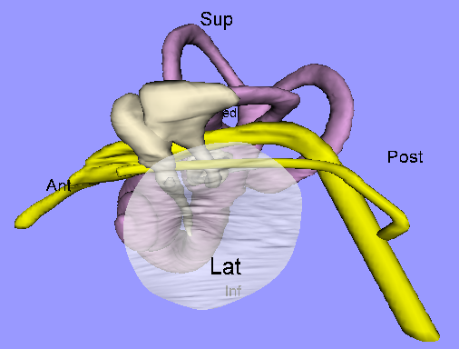
The model shown here includes the middle and inner ear. The grey
part includes the eardrum (translucent) and the three small middle-ear
bones (or ossicles). The mauve part includes the hearing and
balance parts of the inner ear. The yellow structures are nerves.
If you have a VRML viewer you can interact with the model: zoom in and out, rotate in any direction, and see the labels for structures you point at. For further information, and to interact with this model, please see our 3Dear site.
The model is based on a human
magnetic-resonance dataset
provided by Drs. M.M. and O.W. Henson (UNC-Chapel Hill) and the
Center for In
Vivo Microscopy (CIVM) at Duke University.