 Structure and function of the middle ear
Structure and function of the middle ear
Structure and function of the middle ear
After Cull (1989): The Sourcebook of medical illustration, Parthenon, Carnforth, xxiii+481 pp.

After Cull (1989): The Sourcebook of medical illustration, Parthenon, Carnforth, xxiii+481 pp.
After Deaver JB (1900): Surgical anatomy, Vol. 2,
Blakiston, Philadelphia, 709 pp.
Daren Nicholson's 3D
Ear site

Although the configurations are different, in many species there is a second cavity which communicates, through a relatively narrow opening, with the main middle-ear cavity.
This configuration leads to an acoustic
resonance, like a
Helmholtz resonator.
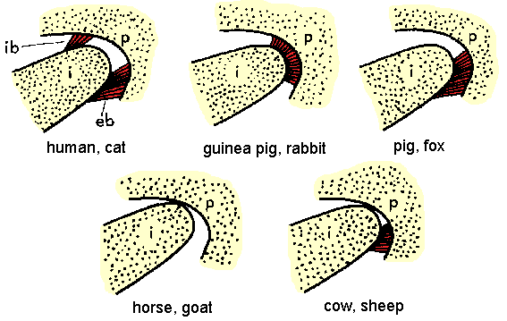
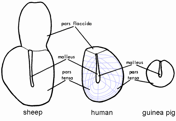 Varying size of pars flaccida.
Sheep after Lim,
Acta Otolaryngol. 66: 515–532 (1968);
human after Filogamo,
Acta Anat. 7: 248–272 (1949)
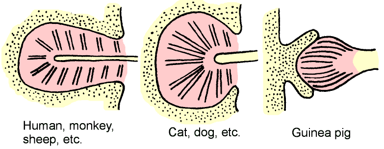
 Varying orientation of manubrium, and varying degrees of
asymmetry.
Varying orientation of manubrium, and varying degrees of
asymmetry.
(Decraemer & Funnell, 2008)

After Gates GR, Saunders JC, Bock GR, Aitkin LM & Elliott MA (1974): Peripheral auditory function in the platypus, Ornithorhynchus anatinus. J Acoust Soc Am 56: 152-156
(not to scale)
Human after Nager GT & Nager M (1953):
The arteries of the human middle ear, with particular regard to
the blood supply of the auditory ossicles.
Ann. Otol. Rhinol. Laryngol. 62: 923-949.
Cat after Jayne H (1898):
Mammalian Anatomy. I. The skeleton of the cat.
Lippincott, Philadelphia, xix+816 pp.

Maftoon et al. (2015): Finite-element modelling of the response of the gerbil middle ear to sound. JARO 16(5): 547–567
Different configurations of posterior incudal ligament in different species.
Based on descriptions by
Kobayashi M (1955a):
On the ligaments and articulations of the auditory ossicles of cow, swine
and goat.
Hiroshima J Med Sci 3: 331-342
Kobayashi M (1955b):
On the ligaments and articulations of the auditory ossicles of the rat
and the guinea pig.
Hiroshima J Med Sci 3: 343-351
Kobayashi M (1955c):
The articulations of the auditory ossicles and their ligaments
of various species of mammalian animals.
Hiroshima J Med Sci 4: 319-349

After Fumagalli (1949): Sound-conducting apparatus: a study of morphology. Arch Ital Otol Rinol e Laringol 60 Suppl. 1: ix+323 pp.
Complex fibre arrangements within ligaments.
After Kobayashi M (1956): The comparative anatomical study of the stapedial muscles of the various kinds of mammalian animals. Hiroshima J Med Sci 5: 63-84
Stapedius muscle in various species

After Fowler EP Jr. (1947): Medicine of the ear, 2nd ed., T. Nelson, New York)
Eardrum becomes more vertical with age.

After Fowler EP Jr. (1947): Medicine of the ear, 2nd ed., T. Nelson, New York)
Fowler (1947): A large group led by Schwalbe believed that the newborn drum ‘slants much more than in adults’ but a group led by Siebenmann did not agree.
Siebenmann (1897, p. 265):
The angle is only slightly less in newborns, and Schwalbe’s observation
that it is almost horizontal was based on a deception.

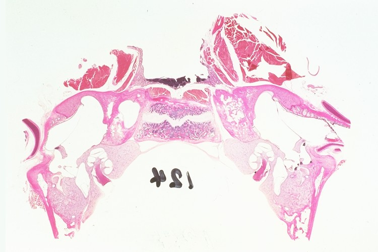
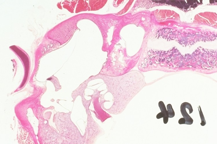
Coronal (frontal) section, reconstructed from horizontal sections from NLM's Visible Human Project.
Click on the image to view a set of images cropped from the original
horizontal sections, in the vicinity of the ear. These are from the
Visible Human female data. The pixel size and slice thickness are
both 0.33 mm.
 Same section, magnified.
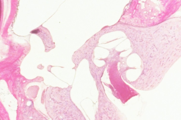


 In a different slice, joint between incus (left) & stapes (right)
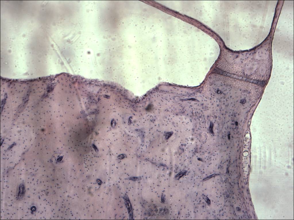
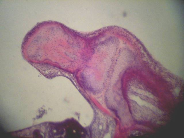
In a different ear
(What's missing in this slide?)


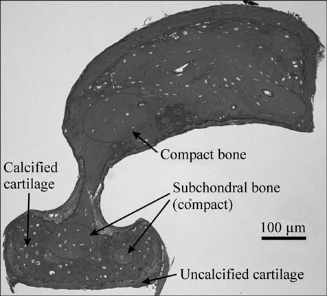
Multiple layers:

Closer.

Closer.
The eardrum is ~10 mm in diameter, but only 10’s of microns thick.

After Fig. 1 in Lim DJ (1968): Tympanic membrane: Electron Microscopic Observation. Part I: Pars Tensa. Acta Oto-Laryngol. 66: 181–198
Three layers:
Layers of lamina propria:
Note the approximately orthogonal fibre organization,
like …
.
Simple model with fixed axis (link to 3-D model).
Matching
low acoustical impedance of air
to
high acoustical impedance of liquid in cochlea.
Mechanisms:
Ratio of eardrum area to footplate area.
Force balance:
| ftm | = | ffp |
| ptmAtm | = | pfpAfp |
| pfp | = | ptm(Atm/Afp) |
After Cull (1989): The Sourcebook of medical illustration, Parthenon, Carnforth, xxiii+481 pp.

Differences in ratios among different families
How to measure the surface areas?
Based on data of Kirikae (1960)
Length of manubrium
vs.
length of long process of incus
Lever arm depends on ...

Simplified model
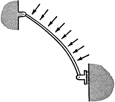
One side only, with distributed load

Further simplification.
Relationship between input xi and output xo?
What assumptions?
Relative magnitudes?
Mechanisms can’t really be separated.
Funnell, J Acoust Soc Am 99: 3036-3043 (1996)

|
Functions:
|
|
Low-frequency measurement with capacitive probe.
 Laser holography.
Simple vibration pattern at low frequencies.
 Literature review shows agreement with Khanna even in older data.
For example, Owada (1959), cat and rabbit
Owada I,
J Otorhinolaryngol Soc Japan 62: 28-43 + 3 plates (1959)
 Kirikae (1960), human
Kirikae I (1960):
The structure and function of the middle ear.
University of Tokyo Press, Tokyo
 Even Békésy's own results can be interpreted as agreeing
in part with Khanna's observations.
Even Békésy's own results can be interpreted as agreeing
in part with Khanna's observations.
 Laser holography.
Vibration pattern breaks up, becomes more complex at high frequencies.
Great variability among individuals.
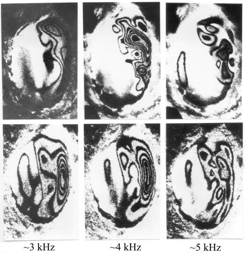
Actual holographic images.
Point-by-point measurements.
Combination of
Measurements required
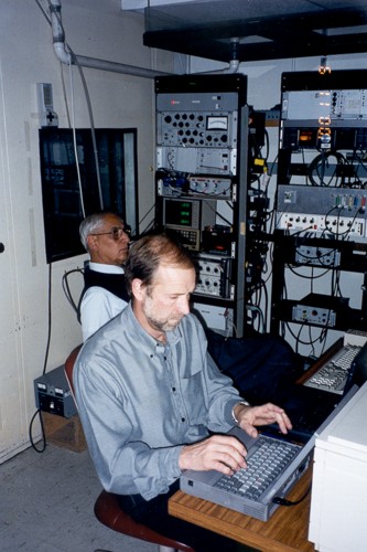
General view of vibration-isolation table inside sound-proof room.
Note nested horizontal and vertical goniometers
on the left.
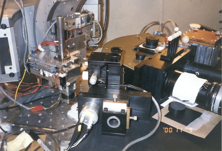
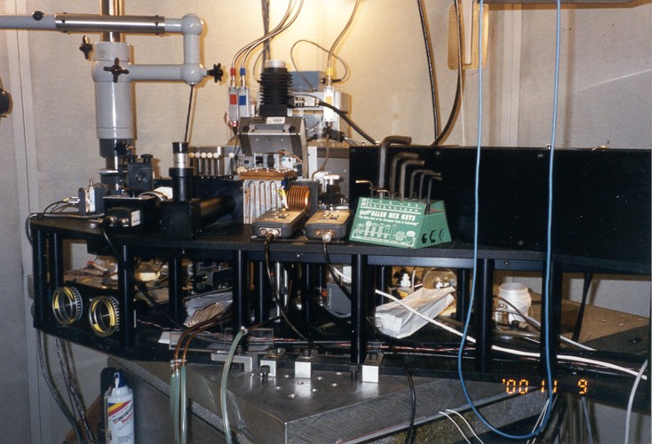
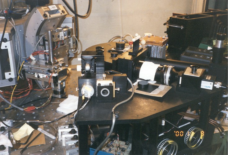
Combined laser interferometer and optical sectioning microscope.
Point-by-point measurements. Cat.
After Fay, Puria, Decraemer & Steele (2005) Fig. 2

Animated point-by-point measurements.
Courtesy W.F. Decraemer

Zinan He (2012), M.Eng. thesis, Mcgill University
Point-by-point measurements. Gerbil.
Off-the-shelf laser Doppler vibrometer designed for clinical use.

Maftoon N, Funnell WRJ, Daniel SJ & Decraemer WF (2013): Experimental study of vibrations of gerbil tympanic membrane with closed middle ear cavity. JARO 14(4): 467-481 (doi:10.1007/s10162-013-0389-9)
Ear canal removed, acoustic coupler attached.

Maftoon N, Funnell WRJ, Daniel SJ & Decraemer WF (2013): Experimental study of vibrations of gerbil tympanic membrane with closed middle ear cavity. JARO 14(4): 467-481 (doi:10.1007/s10162-013-0389-9)
Glass-coated plastic microspheres as laser targets.
Videos of experimental procedure
He Z (2012): Vibration measurements on the widely exposed gerbil eardrum. M.Eng. thesis, McGill University

Maftoon N, Funnell WRJ, Daniel SJ & Decraemer WF (2013): Experimental study of vibrations of gerbil tympanic membrane with closed middle ear cavity. JARO 14(4): 467-481 (doi:10.1007/s10162-013-0389-9)
Vibrations of points on the pars tensa:
Lower inset shows Bode plot, used to confirm phase unwrapping.

Looking into middle ear through hole drilled in bulla.
The manubrium is barely visible.
Note the moist cotton wool and paper towel.

From a slightly different angle, the eardrum and more of the manubrium are visible.
With sufficient precision, vibrations along 3 axes can be measured.

Close-up. The head of the stapes is barely visible at the back.

Close-up from other side, showing the long process of the incus and the
top of the stapes.

Animation showing complex motion of the ossicular chain, as estimated from measurements at multiple points and from multiple directions.
Cf. simple model.

Kose et al. (2019): Vibration measurements of the gerbil eardrum under quasi-static pressure steps
Vibrations measured in the presence of
static pressures:
Frequence responses for +ve and −ve half-cycles.
Shapiro (2014): An experimental study of vibrations in the gerbil middle ear under static pressure.
Vibrations measured in the presence of
static pressures:
3 cycles of pressurization (red, green & blue).
Kose et al. (2021): Vibration measurements of the gerbil eardrum under quasi-static pressure sweeps
Vibrations measured in the presence of
static pressure sweeps: spectrogram of vibrations on manubrium
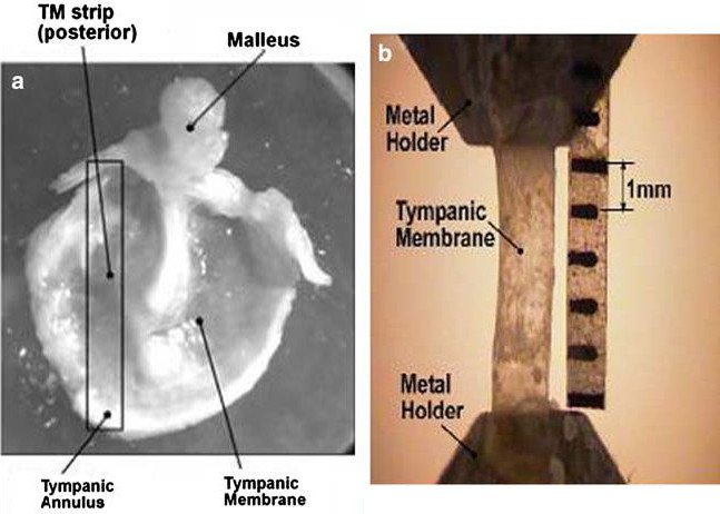
 Transverse
A calibrated hair was used to produce a known bending force on a flap cut from the eardrum.
Békésy, Gv (1949):
The structure of the middle ear and the hearing
of one's own voice by bone conduction.
J Acoust Soc Am 21: 217-232
 For a
calf eardrum. Led to a very low value.
For a
calf eardrum. Led to a very low value.
 Longitudinal
Strip 10 × 1.5 mm.
Vibrator (cantilever beam, natural frequency of 890 Hz).
When the strip of eardrum was attached
to the beam and stretched by a mass, the natural frequency changed.
Kirikae I (1960):
The structure and function of the middle ear.
University of Tokyo Press, Tokyo

Longitudinal
Measured properties as a function of frequency.
Longitudinal
Off-the-shelf instrument
Annals of Biomedical Engineering
35(2): 305–314.
DOI: 10.1007/s10439-006-9227-0
 Great variability between ears.
Voss SE, Rosowski JJ, Merchant SN & Peake WT (2000):
Acoustic responses of the human middle ear.
Hearing Research 150(1-2): 43–69
 One problem is drying.
Ellaham NN, Akache F, Funnell WRJ & Daniel SJ (2007):
Experimental study of the effects of drying
on middle-ear vibrations in the gerbil.
Proc 30th Ann Conf Can Med Biol Eng Soc, paper M0173, 4 pp. (CD-ROM)
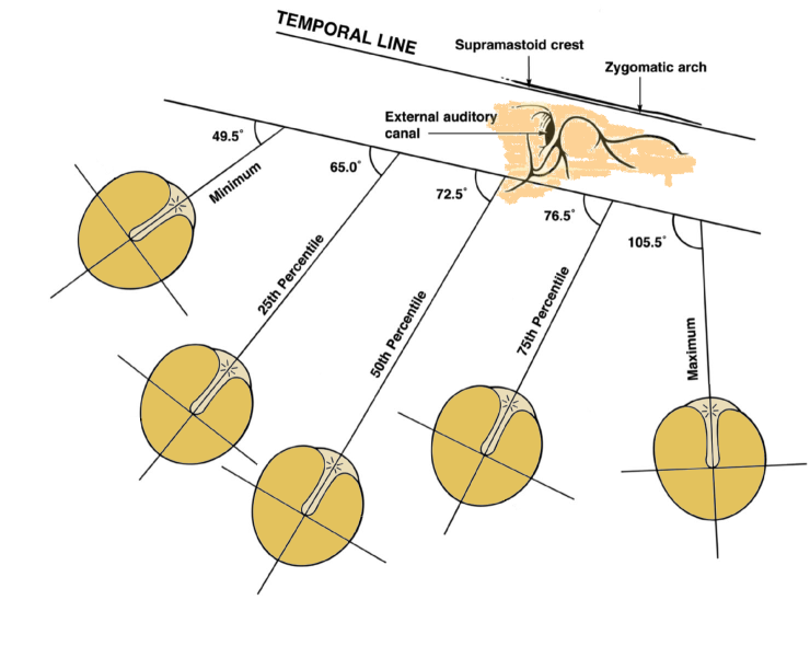
Todd W (2005): Orientation of the manubrium mallei: Inexplicably widely variable. Laryngoscope 115: 1548-1552
Anatomical variability.
For example, orientation of manubrium in human.
Todd NW (2005): Orientation of the manubrium mallei:
Inexplicably widely variable. Laryngoscope 115:
1548–1552
BMDE-501
Modelling
middle-ear mechanics
