Creation of 3-D models
Creation of 3-D models
Creation of 3-D models
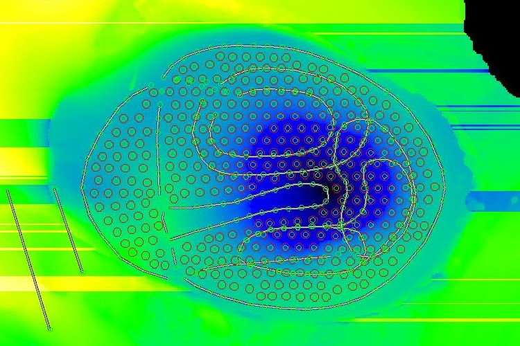
Decraemer and Dirckx in Antwerp can measure the precise 3-D shape of the eardrum using phase-shift moiré topography, a non-contacting optical method.
The method uses a grid of straight lines.
A light shone through the grid onto a curved surface causes curved shadows.
Looking at the shadows through the grid produces a moiré interference pattern.
The illusory lines of the interference pattern form contours of constant depth, defining the shape of the surface.
We obtain an image of the eardrum in which the grey level of each pixel
is proportional to the z coördinate (modulo 2π times a
constant).
The application of static pressures during the shape measurement allows us to
Special software allows us to transfer the z
coördinates from the moiré image to a finite-element
mesh.
Principle of tomographic reconstruction: simple back-projection algorithm

Animation produced by bapr.
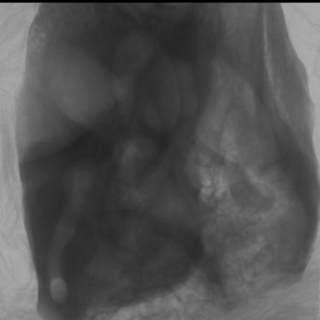
Single microscopic X-ray image of human middle and inner ear.
Taken with SkyScan 1072 scanner.
Resolution down to 5 μm if specimen small enough.
Mainly bone.
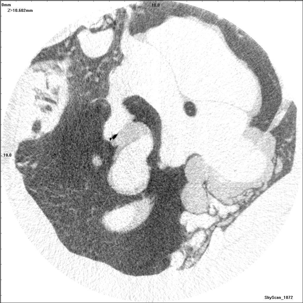
μCT reconstructed slice (human) showing eardrum and manubrium.
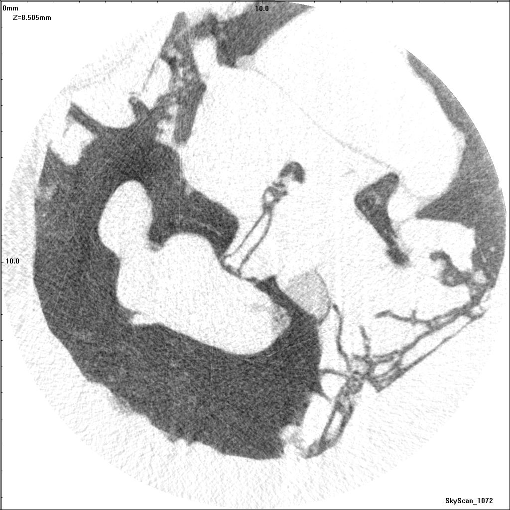
μCT reconstructed slice showing stapes and tip of incus.
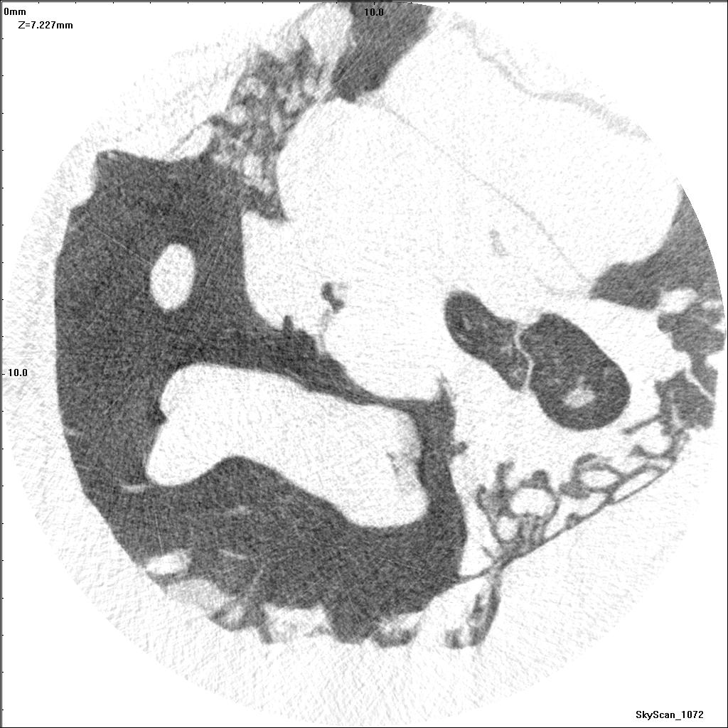
μCT reconstructed slice showing heads of malleus and incus.
3-D volume.

Soft tissues more clearly visible.
Resolutions down to 10’s of μm.

Great detail; problems with distortion and alignment

Expensive and time-consuming,
especially for 1-µm sections
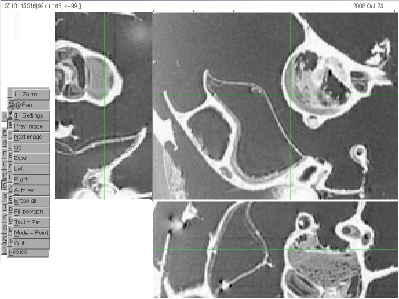
Image segmentation with stacks of images.
Threshold-based segmentation
Fast but often unsatisfying.

Good for some applications.

Not so good for complex models.
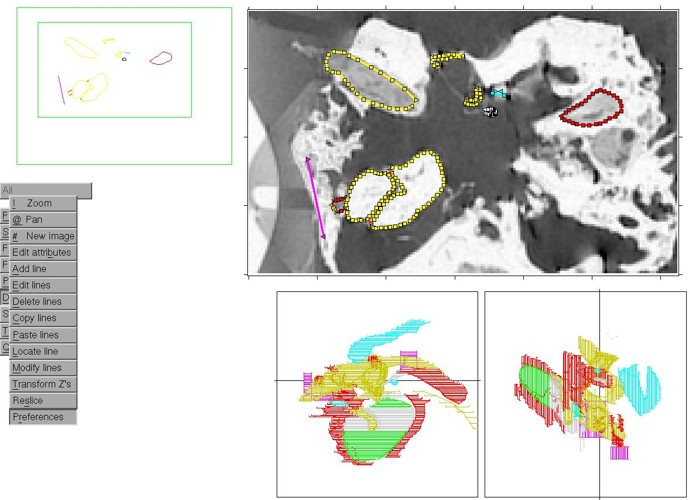
Manual segmentation.

Semi-automatic segmentation with ‘snake’ algorithm
Triangulation across slices to form surface

Complex and irregular surfaces

Connections and shared surfaces
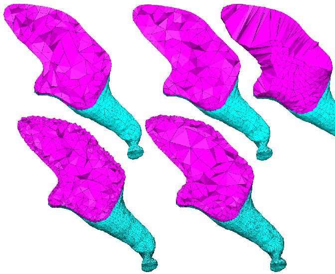
Volume meshes
Variety of algorithms
BMDE-501
Modelling
middle-ear mechanics