
An existing finite-element model of the cat middle ear was modified to include a shell representation of the incudostapedial joint. Joint stiffness was varied from very small to very large and the results show that both out-of-plane and in-plane displacements become smaller as the joint becomes more flexible. In-plane rotations remain significant at all stiffness values and tilting varies considerably in magnitude and direction as a function of stiffness.Introduction
The finite-element method has been used to model both cat [1], [2] and human eardrums [3]. More recently, these models have been extended to include the ossicles and cochlear load [4], [5]. The incudostapedial joint was taken to be rigid in these models.
Experimental results indicate that footplate motion is primarily piston-like, but rigid-joint models show footplate tilting and in-plane displacements. There is some experimental evidence that the incudostapedial (i-s) joint is not rigid [6]. In this paper, we introduce a flexible i-s joint into our finite-element model of the cat middle ear.
Methods
The model presented here is identical to our earlier model [5] except for the explicit representation of the i-s joint. Twelve shell elements with a thickness of 0.15 cm were used to model the surface of the i-s joint. These elements are not intended to realistically represent the detailed anatomy of the joint but provide a preliminary implementation of joint flexibility.
The Young's modulus of the elements representing the i-s joint was varied from 2 * 10^5 to 2 * 10^11 dyn cm^-2 to examine the effects of joint flexibility. The simulated input to the model was a uniform static pressure on the eardrum. The x, y, and z displacements of an anterior and a posterior node of the footplate were used to characterize footplate displacements, including both translational and rotational movement.
Results
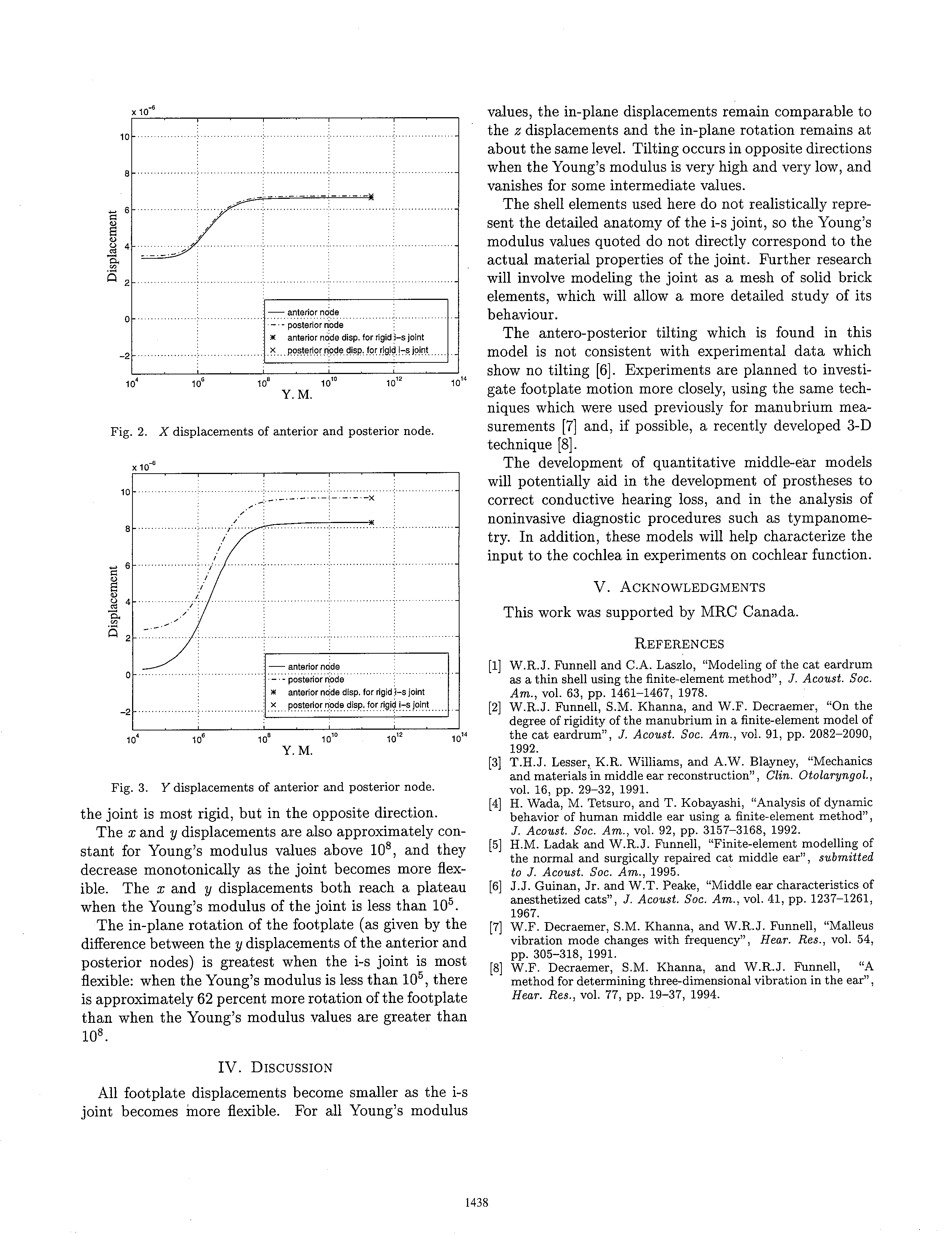
Figures 1, 2, and 3 show the displacements along the z, x, and y directions, respectively, of the anterior and posterior nodes, as functions of the Young's modulus of the i-s joint. The '*' and 'x' symbols represent the displacements obtained for a finite-element model in which the i-s joint was perfectly rigid [5]. The footplate lies in the x-y plane, with the x-axis running antero-posteriorly. Thus, both the x and y displacements of the anterior and posterior nodes are in plane displacements while the z displacement is the out-of-plane displacement. The z displacement is the most important component of footplate displacement because it is the effective input to the fluid of the cochlea. The difference between the z displacements of the anterior and posterior nodes is proportional to the tilting of the footplate about the y axis.
[Figure 1 omitted]
[Figure 2 omitted]
[Figure 3 omitted]
Figure 1 shows that the z displacements are approximately constant for Young's modulus values above 10^8, and then generally decrease as the Young's modulus decreases. When the Young's modulus is high, the displacement of the anterior node is 20 percent larger than that of the posterior node, indicating significant antero-posterior tilting. At a Young's modulus of approximately 6 * 10^6, the anterior and posterior z displacements are equal, meaning that there is no antero-posterior tilting of the footplate. At this point the two displacement curves cross and the footplate starts to tilt in the opposite direction. When the i-s joint is most flexible, the amount of antero-posterior tilting is 3.5 times greater than when the joint is most rigid, but in the opposite direction.
The x and y displacements are also approximately constant for Young's modulus values above 10^8, and they decrease monotonically as the joint becomes more flexible. The x and y displacements both reach a plateau when the Young's modulus of the joint is less than 10^5.
The in-plane rotation of the footplate (as given by the difference between the y displacements of the anterior and posterior nodes) is greatest when the i-s joint is most flexible: when the Young's modulus is less than 10^5, there is approximately 62 percent more rotation of the footplate than when the Young's modulus values are greater than 10^8.
Discussion
All footplate displacements become smaller as the i-s joint becomes more flexible. For all Young's modulus values, the in-plane displacements remain comparable to the z displacements and the in-plane rotation remains at about the same level. Tilting occurs in opposite directions when the Young's modulus is very high and very low, and vanishes for some intermediate values.
The shell elements used here do not realistically represent the detailed anatomy of the i-s joint, so the Young's modulus values quoted do not directly correspond to the actual material properties of the joint. Further research will involve modeling the joint as a mesh of solid brick elements, which will allow a more detailed study of its behaviour.
The antero-posterior tilting which is found in this model is not consistent with experimental data which show no tilting [6]. Experiments are planned to investigate footplate motion more closely, using the same techniques which were used previously for manubrium measurements [7] and, if possible, a recently developed 3-D technique [8].
The development of quantitative middle-ear models will potentially aid in the development of prostheses to correct conductive hearing loss, and in the analysis of noninvasive diagnostic procedures such as tympanometry. In addition, these models will help characterize the input to the cochlea in experiments on cochlear function.
Acknowledgments
This work was supported by MRC Canada.
References
Funnell, W.R.J. and Laszlo, C.A., "Modeling of the cat eardrum as a thin shell using the finite-element method", J. Acoust. Soc. Am., vol. 63, pp.1461-1467, 1978.
Funnell, W.R.J. and Khanna, S.M. and Decraemer, W.F., "On the degree of rigidity of the manubrium in a finite-element model of the cat eardrum", J. Acoust. Soc. Am., vol. 91, pp. 2082-2090, 1992.
Lesser, T.H.J. and Williams, K.R. and Blayney, A.W., "Mechanics and materials in middle ear reconstruction", Clin. Otolaryngol., vol. 16, pp. 29-32, 1991.
Wada, H. and Tetsuro, M. and Kobayashi, T., "Analysis of dynamic behavior of human middle ear using a finite-element method", J. Acoust. Soc. Am., vol. 92, pp. 3157-3168, 1992.
Ladak, H.M. and Funnell, W.R.J., "Finite-element modelling of the normal and surgically repaired cat middle ear", submitted to J. Acoust. Soc. Am., 1995.
Guinan, Jr., J.J. and Peake, W.T., "Middle ear characteristics of anesthetized cats", J. Acoust. Soc. Am., vol. 41, pp. 1237-1261, 1967.
Decraemer, W.F. and Khanna, S.M. and Funnell, W.R.J., "Malleus vibration mode changes with frequency", Hear. Res., vol. 54, pp. 305-318, 1991.
Decraemer, W.F. and Khanna, S.M. and Funnell, W.R.J., "A method for determining three-dimensional vibration in the ear", Hear. Res., vol. 77, pp. 19-37, 1994.